2048
Magnetization Transfer Contribution to Balanced SSFP Image Contrast in Chronic Myocardial Infarction1Bioengineering Department, University of California, Los Angeles, CA, United States, 2Biomedical Imaging Research Institute, Cedars-Sinai Medical Center, Los Angeles, CA, United States, 3Department of Medicine, University of California, Los Angeles, CA, United States
Synopsis
Magnetization Transfer (MT) effect in balanced steady-state free precession (bSSFP) has been demonstrated in acute myocardial infarction (MI). However, whether MT effect influences image contrast in bSSFP acquisitions of chronic MI has not been investigated. We studied this using a collagen phantom and large animal models with chronic MI. We found the bSSFP MT ratio to be more than 2-fold higher in the chronic MI zone compared to uninfarcted myocardium. Our findings support the notion that a strong MT contrast in bSSFP acquisitions, likely from collagen deposition within chronic MI, is available for discriminating chronic MI territories without contrast agents.
Introduction
The influence of Magnetization Transfer (MT) effect in balanced steady-state free precession (cine-bSSFP) has been demonstrated in acute myocardial infarction (MI).1 To study this, previous studies used dual bSSFP acquisitions - one performed with a RF duration imposing a high MT effect and another that is minimally sensitive to MT effect.2 However, whether MT contrast influences image contrast in bSSFP acquisitions in the setting of chronic MI, where the tissue characteristics are fundamentally different compared to acute MI, has not been investigated. Given chronic MI regions have extensive collagen deposition (and hence large macromolecular pool), we hypothesized that MT contrast may be also be observable in bSSFP acquisitions. We tested our hypothesis using a collagen phantom and large animal models with chronic MI.Methods
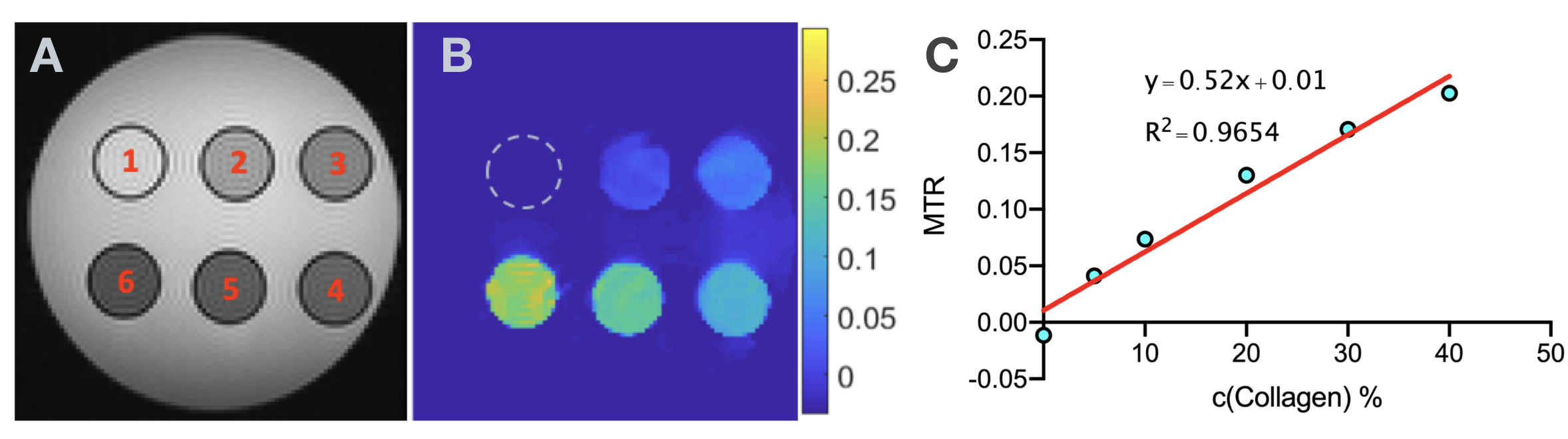
Phantom Preparation: To investigate the MT contribution from collagen in bSSFP sequences, we performed a phantom study with six tubes containing Type I and Type III collagen (NeoCell, Newport Beach, CA) at 0%, 5%, 10%, 20%, 30% and 40% concentration in distilled water, which were arranged in a container filled with water (Fig. 1A) for imaging.Animal Preparation: To examine the MT effects in vivo, dogs (n = 7; 20-30 kg body weight) were studied according to the protocols approved by Institutional Animal Care and Use Committees. Myocardial infarction was created in dogs by ligating the left anterior descending (LAD) artery for 2 hours followed by reperfusion. The animals were allowed to recover and studied at least 6 months after the induction of MI.
Image acquisition and analysis: All images were acquired in 3.0T MRI system (Biograph mMR, Siemens Healthcare, Erlangen, Germany). Balanced SSFP images were acquired with continuous RF excitations with the following scan parameters: flip angle = 45°, bandwidth = 1002 Hz/pixel, voxel size=1.6 x 1.6 x 6 mm3, retrospective ECG gating with variable RF duration (τRF) = [800, 2000] ms along with variable TR (3.18, 4.38 ms respectively) were acquired in dogs with MI. LGE images (IR-prepared FLASH; TI optimized to null remote myocardium; TR/TE=3.5/1.75ms) with the same resolution were acquired at the end of the scan for comparison. MT Ratio (MTR) map was computed as
$$MTR = \frac{S_{TR=4.38 ms} - S_{TR=3.18 ms}}{S_{TR=4.38 ms}}$$ where $$$S_{TR=4.38 ms}$$$ and $$$S_{TR=3.18 ms}$$$ denote bSSFP signal from acquisitions with TR = 4.38 ms and 3.18 ms respectively. ROIs in MTR map were manually drawn according to the ROIs in LGE. The distribution of MTR between MI and remote myocardium were compared using pairwise t-test and percentage difference was also calculated.
Results
There was a strong relationship between bSSFP MTR and collagen concentration in in-vitro preparation (Fig. 1B - 1C). As shown in Fig. 2, there was high MTR contrast between chronic MI region and remote myocardium in MTR maps with excellent spatial correspondence with the chronic MI territories identified on LGE (ref ROI in Fig. 2A). bSSFP MTR for the MI region was 0.43 ± 0.11, which was significantly higher (115%) than the bSSFP MTR for remote myocardium (0.20 ± 0.04, Fig. 2C).Discussion
We found that MT contribution to bSSFP signal is highly sensitive to collagen concentration in vitro and that the bSSFP MTR is more than 2-fold higher in the chronic MI zone compared to remote/uninfarcted myocardium. Additional studies are needed to fully understand the biophysical mechanisms contributing to the MT-based image contrast in chronic MI.Conclusion
A strong MT contrast in bSSFP acquisitions, likely from collagen deposition within chronic MI, is available for discriminating chronic MI territories without contrast agents at 3T.Acknowledgements
No acknowledgement found.References
1. Weber OM, Speier P, Scheffler K, Bieri O. Assessment of magnetization transfer effects in myocardial tissue using balanced steady-state free precession (bSSFP) cine MRI. Magn Reson Med. 2009;62(3):699-705.
2. Bieri O, Scheffler K. Optimized balanced steady-state free precession magnetization transfer imaging. Magn Reson Med. 2007;58(3):511-8.
Figures