2046
Model-based reconstruction for simultaneous multi-slice myocardial T1 mapping using single-shot inversion-recovery radial FLASH1Department of Interventional and Diagnostic Radiology of the University Medical Center Göttingen, Göttingen, Germany, 2DZHK (German Centre for Cardiovascular Research), partner site Göttingen, Göttingen, Germany
Synopsis
Fast multi-slice myocardial T1 mapping is of great interest in clinical cardiovascular magnetic resonance (CMR) imaging. This work extends a simultaneous-multi-slice (SMS) model-based reconstruction method for 3-slice myocardial T1 mapping using single-shot Inversion-recovery radial FLASH. Initial results on experimental phantom and one healthy volunteer have demonstrated simultaneous 3-slice myocardial T1 mapping (1.6 x 1.6 x 8 mm3 ) might be feasible within a single inversion recovery of 4 s. More clinical validations of the proposed method will be explored in future studies.
Introduction
Quantitative myocardial T1 mapping is of increasing interest in clinical cardiovascular magnetic resonance (CMR) imaging [1,2]. While conventional MOLLI-based methods use 2 to 3 inversions for data acquisition [3], recent developments in non-Cartesian sampling [4-6] in combination with advanced reconstructions such as compressed sensing [6], model-based reconstructions [7-10] have enabled single-slice myocardial T1 mapping within a single inversion-recovery (IR). To further enable T1 mapping with multiple sections within a single inversion, this work extends a simultaneous-multi-slice (SMS) model-based reconstruction method for high-resolution 3-slice myocardial T1 mapping using single-shot IR radial FLASH. Validations of the proposed method have been performed on an experimental phantom and one healthy subject.Methods
We employ a triggered IR SMS radial FLASH scheme for data acquisition: After a non-selective inversion, radial SMS data is continuously acquired using a golden-angle FLASH readout with the rotated golden-angle used in the partition dimension [11, 12]. To eliminate effects from systolic motion, only data from the diastolic phase is retrospectively selected for T1 estimation. Similar to [11], let $$$p, q$$$ define the partition index and the slice index and let $$$Q$$$ be the total number of partitions/slices. The estimation of parameter maps and coil sensitivity maps from all slices is formulated as a single nonlinear inverse problem [12]:$$\hat{x} = \text{argmin}_{x\in D} \|F(x) -\widetilde{Y} \|_{2}^{2} + \alpha \sum_{q=1}^{Q}R(x^{q}_{p}) + \beta \sum_{q=1}^{Q}U(x^{q}_{c})$$
with $$$F$$$ a nonlinear operator which maps all unknowns x from all slices to the measured undersampled SMS data $$$\widetilde{Y}$$$. $$$x^{q}_{p}$$$ contains steady-state signal $$$M_{ss}$$$, equilibrium signal $$$M_{0}$$$ and effective relaxation rate $$$R_{1}^{*}$$$ of the $$$q$$$th slice, while $$$x^{q}_{c}$$$ is a set of the corresponding coil sensitivity maps. $$$R(\cdot)$$$ defines the joint $$$\ell_{1}$$$-Wavelet regularization in the parameter dimension [8], $$$U({\cdot})$$$ represents the Sobolev norm enforcing the smoothness of coil sensitivity maps. $$$\alpha$$$ and $$$\beta$$$ are the regularization parameters for parameter maps and coil sensitivity maps, respectively. $$$D$$$ is a convex set ensuring the effective relaxation rate $$$R_{1}^{*}$$$ to be nonnegative [8]. The above nonlinear inverse problem is solved by the IRGNM-FISTA algorithm [8].
Data acquisition was performed on a Magnetom Skyra 3T (Siemens Healthineers, Erlangen, Germany). Phantom studies were conducted with a 20-channel head/neck coil, whereas combined thorax and spine coils with 26 channels were utilized for in-vivo scans. The acquisition parameters for phantom were: FOV: 192 x 192 mm$$$^{2}$$$, matrix size: 192 x 192, slice thickness 5 mm (gap 15 mm), TR/TE = 4.10/2.58 ms, bandwidth 630 Hz/pixel. Parameters for the in-vivo measurements (female, age 27, heart rates 55 bpm) were: FOV: 256 x 256 mm$$$^{2}$$$, matrix size: 160 x 160, slice thickness 8 mm (gap 20 mm), TR/TE = 2.90/1.79 ms, bandwidth 850 Hz/pixel. All single-shot measurements were acquired within 4 seconds using the nominal flip angle $$$6^{\circ}$$$. In-vivo experiments were performed during a brief breathhold. All data processing was done offline and implemented in BART [13].
Results
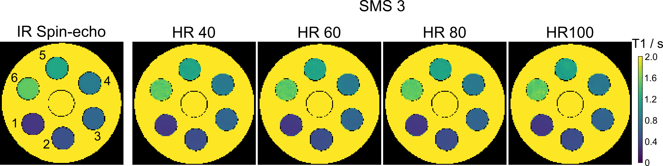
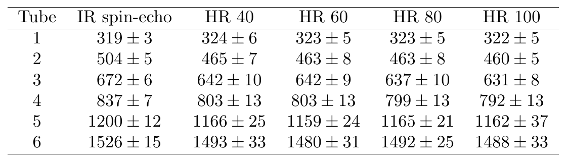
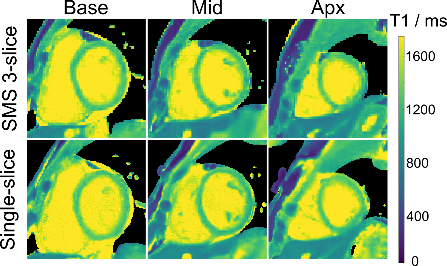
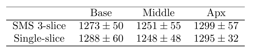
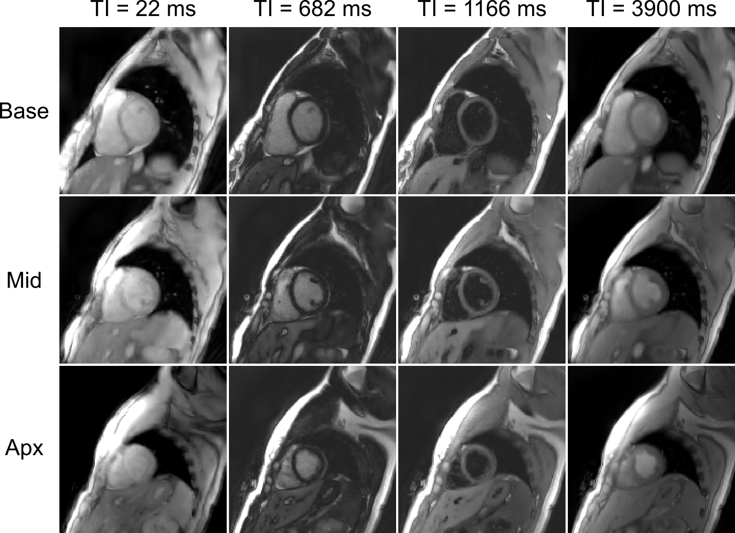
Figure 1 shows the estimated SMS T1 maps (center-slice) of an experimental phantom for simulated heart rates between 40 and 100 bpm. Visual inspection reveals good agreement for all heart rates and T1 values when compared to a conventional IR spin-echo method. These findings are confirmed by quantitative ROI-analyses summarized in Table 1. Figure 2 presents 3-slice myocardial T1 maps acquired using single-shot IR SMS radial FLASH and reconstructed by the proposed model-based algorithm. All 3-slice T1 maps are visually in good agreement with the corresponding single-slice ones, which is further confirmed in the ROI-analyzed septal T1 values in Table 2. Further, figure 3 demonstrates four synthetic T1-weighted images for all 3 slices, which shows a distinct contrast between myocardium and blood pool.Discussion & Conclusion
This work extends IR SMS radial FLASH sequence and sparsity-constrained model-based reconstructions [12] into single-shot multi-slice myocardial T1 mapping. Initial results on phantom and one healthy volunteer have demonstrated simultaneous 3-slice myocardial T1 mapping might be feasible within a single inversion recovery of 4 s. The main limitation of the present work is the limited number of subjects. More clinical validations of the proposed method will be explored in future studies.Acknowledgements
No acknowledgement found.References
1. Moon JC, Messroghli DR, Kellman P, Piechnik SK, Robson MD, Ugander M, Gatehouse PD, Arai AE, Friedrich MG, Neubauer S, et al. Myocardial T1 mapping and extracellular volume quantification: a society for cardiovascular magnetic resonance (SCMR) and CMR working group of the European society of cardiology consensus statement. J Cardiovasc Magn Reson. 2013;15:92.
2. Kellman P, Hansen MS. T1-mapping in the heart: accuracy and precision. J Cardiovasc Magn Reson. 2014;16:2.
3. Messroghli DR, Radjenovic A, Kozerke S, Higgins DM, Sivananthan MU, Ridgway JP. Modified look-locker inversion recovery (MOLLI) for high-resolution T1 mapping of the heart. Magn Reson Med. 2004;52:141–6.
4. Gensler D, Mörchel P, Fidler F, Ritter O, Quick HH, Ladd ME, Bauer WR, Ertl G, Jakob PM, Nordbeck P. Myocardial T1: quantification by using an ECG-triggered radial single-shot inversion-recovery MR imaging sequence. Radiology. 2014;274:879–87.
5. Wang X, Joseph AA, Kalentev O, Merboldt KD, Voit D, Roeloffs V, van Zalk M, Frahm J. High-resolution myocardial T1 mapping using single-shot inversion-recovery fast low-angle shot MRI with radial undersampling and iterative reconstruction. Br J Radiol. 2016;89:20160255.
6. Marty B, Coppa B, Carlier P. Fast, precise, and accurate myocardial T1 mapping using a radial MOLLI sequence with FLASH readout. Magn Reson Med. 2018;79:1387–98.
7. Block KT, Uecker M, Frahm J. Model-based iterative reconstruction for radial fast spin-Echo MRI. IEEE Trans Med Imaging. 2009;28:1759–69.
8. Wang X, Roeloffs V, Klosowski J, Tan Z, Voit D, Uecker M, Frahm J. Model-based T1 mapping with sparsity constraints using single-shot inversion-recovery radial FLASH. Magn Reson Med. 2018;79:730–40.
9. Becker KM, Schulz-Menger J, Schaeffter T, Kolbitsch C. Simultaneous high-resolution cardiac T1 mapping and cine imaging using model-based iterative image reconstruction. Magn Reson Med. 2019;81:1080–91.
10. Wang X., Kohler F., Unterberg-Buchwald C. et al. Model-based myocardial T1 mapping with sparsity constraints using single-shot inversion-recovery radial FLASH cardiovascular magnetic resonance. J Cardiovasc Magn Reson 21, 60 (2019) doi:10.1186/s12968-019-0570-3.
11. Rosenzweig S, Holme HCM, Wilke RN, Voit D, Frahm J, Uecker M. Simultaneous multi-slice MRI using cartesian and radial FLASH and regularized nonlinear inversion: SMS-NLINV. Magn Reson Med. 2018;79:2057–66.
12. Wang X, Rosenzweig S, Scholand N, Holme H.C.M., Uecker M. Model-based Reconstruction for Simultaneous Multi-slice T1 Mapping using Single-shot Inversion-recovery Radial FLASH. arXiv:1909.10633.
13. Uecker M, Ong F, Tamir J, Bahri D, Virtue P, Cheng J, Zhang T, Lustig M. Berkeley advanced reconstruction toolbox. In: Proceedings of the 23rd annual meeting of ISMRM. Toronto; 2015. p. 2486.
Figures