2041
Changes in cerebral venous blood oxygen levels in patients with end-stage renal disease undergoing hemodialysis1Department of Radiology, The First Affiliated Hospital of Dalian Medical University, Dalian, China, 2Philips Healthcare, Beijing, China
Synopsis
End-stage renal disease (ESRD) usually requires maintenance dialysis or kidney transplantation. However, whether maintenance of hemodialysis improves cerebral oxygen levels in patients with ESRD is unclear. This study aim to explore the changes in blood oxygen content in the venules of patients with ESRD who maintained hemodialysis were evaluated by measuring the phase value (Δφ) of deep venous magnetic susceptibility (SWI) in the deep brain. We found that the cerebral venous oxygen level in patients with hemodialysis ESRD was higher than that in the healthy control group. Maintaining hemodialysis may improve cerebral blood oxygen levels in ESRD patients.
Introduction
End-stage renal disease (ESRD) has become a worldwide public health problem, and it is a risk factor for vascular disease, stroke, and cognitive dysfunction. ESRD results in excessive accumulation of urea and toxic metabolites. Moreover, maintenance hemodialysis or kidney transplantation is usually performed in patients with ESRD. However, whether maintenance of hemodialysis improves cerebral oxygen levels in patients with ESRD is unclear. In recent years, the application of magnetic sensitive weighted imaging (SWI) in the diagnosis of central nervous system lesions has attracted more and more attention. The phase (Δφ) of the structure in the brain can be measured by the SWI phase diagram1, while the venous blood oxygen content is the basis of SWI angiography, so measuring vein Δφ can reflect venous blood oxygen levels. The following study aim to explore the changes in blood oxygen content in the venous venules of patients with ESRD who maintained hemodialysis were evaluated by measuring the Δφ of deep venous magnetic susceptibility (SWI) in the deep brain.Materials and Methods
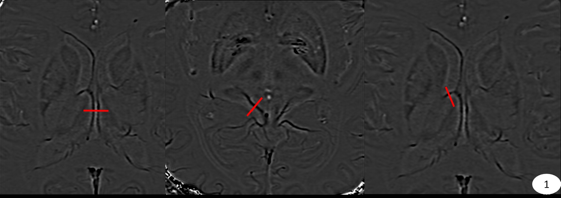
Fourteen maintenance hemodialysis ESRD patients(8 males,6 females,59.7 ± 11.7 yrs) and 13 age- and sex-matched healthy volunteers (6 male,7 female, age 52.5 ± 10.0 yrs) were participated in this retrospective study. All subjects underwent SWI on a 3.0 T MR scanner (Ingenia CX, Philips Healthcare, Best, the Netherlands) with a 32-channel neck-head array coil. And the processed SWI phase map was imported into a personal computer. The SPIN software was used to measure the bilateral internal cerebral vein (ICV) and mound,superior thalamostriate vein (STV), basal vein (BV), manually draw the vertical line of the vein, through the vein and surrounding parenchyma (Fig.1). The software automatically generateed a measured value curve, then the phase difference Δφ between the vein and the surrounding tissue was calculated. The phase difference between the vein and the surrounding tissue was linear with the blood oxygen saturation Y of the vein. Therefore, the blood oxygen content of the brain vein was estimated by Δφ. Using SPSS software, the difference between ESRD group and control group was analyzed by T test. The correlation between the phase value of each vein in ESRD group and the duration of dialysis was tested by Pearson correlation test.Results
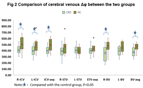
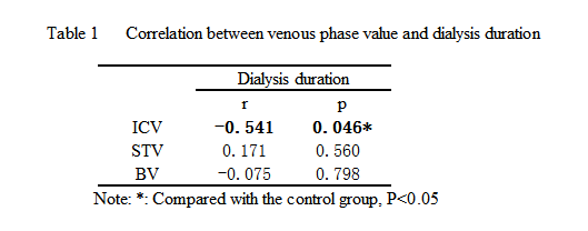
The bilateral ICVΔφ of the ESRD group (408.35±70.29, 392.70±76.39) were significantly lower than that of the control group (501.03±120.28, 483.74±115.45); the right BVΔφ of the ESRD group (353.93±121.79) was lower than that of the control group (467.20 ±107.66) (P<0.05); the averaged Δφ of ICV, and BV in the ESRD group(400.52±65.37,375.67±79.12) were also significantly lower than those in the control group(492.39±99.08,461.91±59.45), but there was no significant difference in BTVΔφ.(Fig.2) There was no difference in the bilateral Δφ of each vein in the two groups. The averaged ICVΔφ in ESRD patients was inversely correlated with the duration of patient dialysis(Table 1).Discussion and Conclusion
SWI venous structural imaging relies on both the shortening of the T2* time caused by the inhomogeneous magnetic field induced by deoxyhemoglobin and the phase difference between the vein and the surrounding tissue. Hemoglobin has two states of oxygenated hemoglobin and deoxyhemoglobin in the human body, and the magnetic susceptibility of the two is different. Oxygenated hemoglobin has a very low antimagnetic rate relative to surrounding brain tissues, and deoxyhemoglobin is paramagnetic with respect to surrounding brain tissues. Therefore, changes in oxygen saturation cause a phase difference between the vein and the surrounding tissue2.3. In this study, the bilateral ICVΔφ, the right BV Δφ, and the average Δφ of ICV and BV in the ESRD group were lower than those in the control group, suggesting that the cerebral venous oxygen level in patients with dialysis ESRD was higher than that in the healthy control group. It indicated that dialysis may improve the blood oxygen level in the brain of ESRD patients. And the longer the regular dialysis duration, the better the improvement of intracerebral venous blood oxygen level, suggesting that the regular maintenance of the dialysis room in ESRD patients may improve the blood oxygenation in the brain.So, Maintaining hemodialysis may improve cerebral blood oxygen levels in ESRD patients.Acknowledgements
No acknowledgement found.References
1.Li M, Hu J, Miao Y, et al. In vivo measurement of oxygenation changes after stroke using susceptibility weighted imaging filtered phase data[J]. PloS one, 2013, 8(5): e63013.
2. Schaller B,Graf R.Cerebral venous infarction:the pathophysiological concept.Cerebrovasc Dis,2004,18 (3):179-188.
3.Schmierer K,Parkes HG,So PW,et al.High field(9.4 Tesla)magnetic resonance imaging of cortical grey matter lesions in multiple sclerosis.Brain,2010,133(Pt 3):858-867.
Figures