2039
Effect of Jet Lag on Brain White Matter Functional Connectivity1Huaxi MR Research Center (HMRRC), Department of Radiology, West China Hospital of Sichuan University, Chengdu 610041, China, ChengDu, China
Synopsis
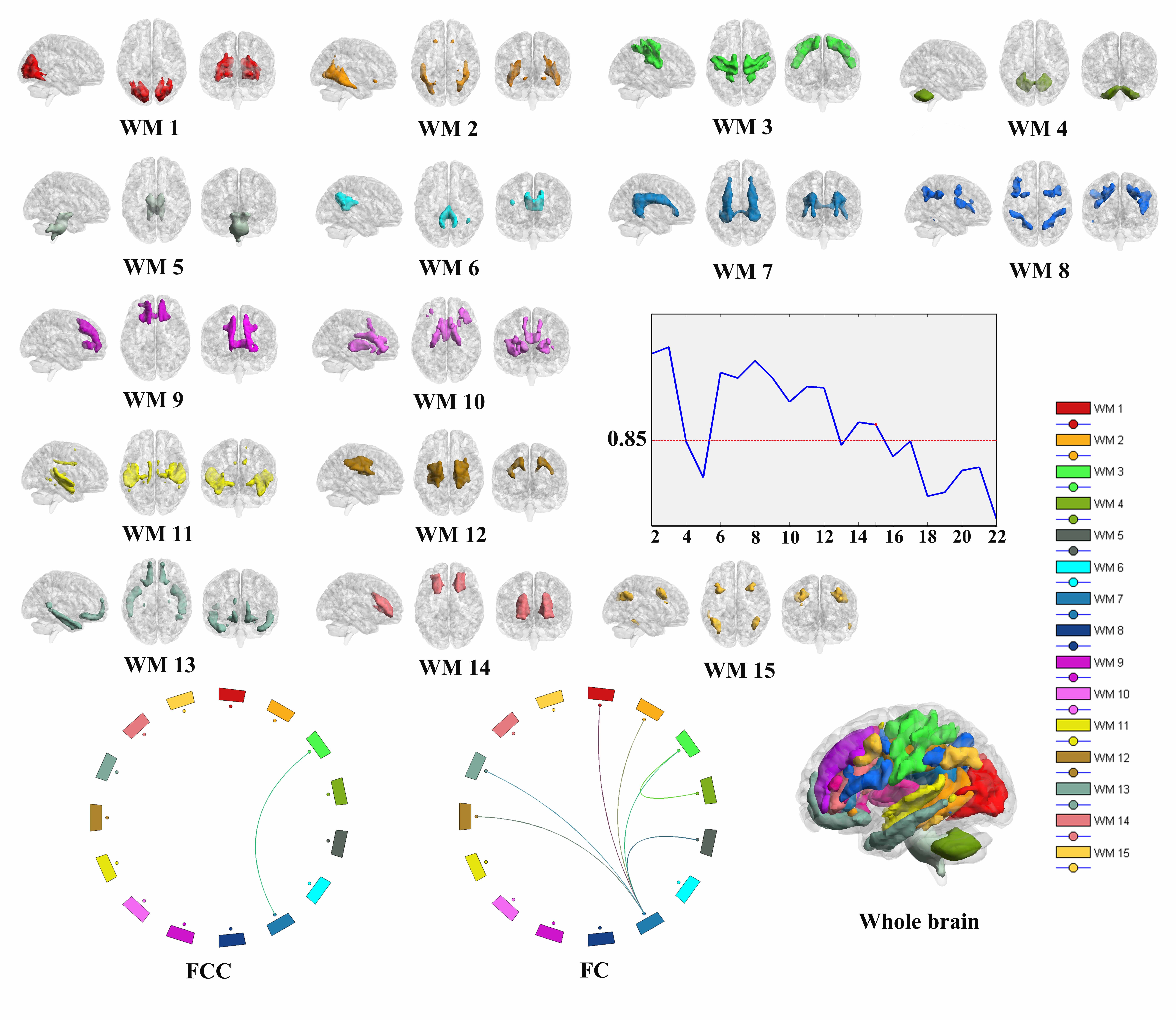
The most prominent finding is decreased static FC and FCC between the cingulate network (WM7) and other WM networks, especially the occipital network, precentral/postcentral network, inferior longitudinal fasciculus network and corona radiate network. The cingulate network is also identified as being affected by Jet Lag in that there is a significant negative correlation between the cingulate and WM3 and GM7 and positive emotion scores. Taken together, these findings provide evidence that changes in WM FC, and especially of the cingulate, may be linked to an decrease in positive emotion as a symptom of Jet Lag.
Background
The circadian rhythms of a person (commonly referred to as the “body” clock) are generally synchronous with the light-dark solar cycle [1]. A long haul flight across more than five time zones may, however, break this balance and produce a circadian rhythm disorder known as Jet Lag [2], the main manifestation of which is excessive daytime sleepiness [3], and additional symptoms may include altered mood [4], gastrointestinal distress, fatigue and cognitive impairments such as memory and concentration deficits [5-7]. Jet Lag has been reported to be associated with alterations of brain Functional Connectivity (FC) in gray matter. However, little is known about the effect of Jet Lag on white matter FC.Methods
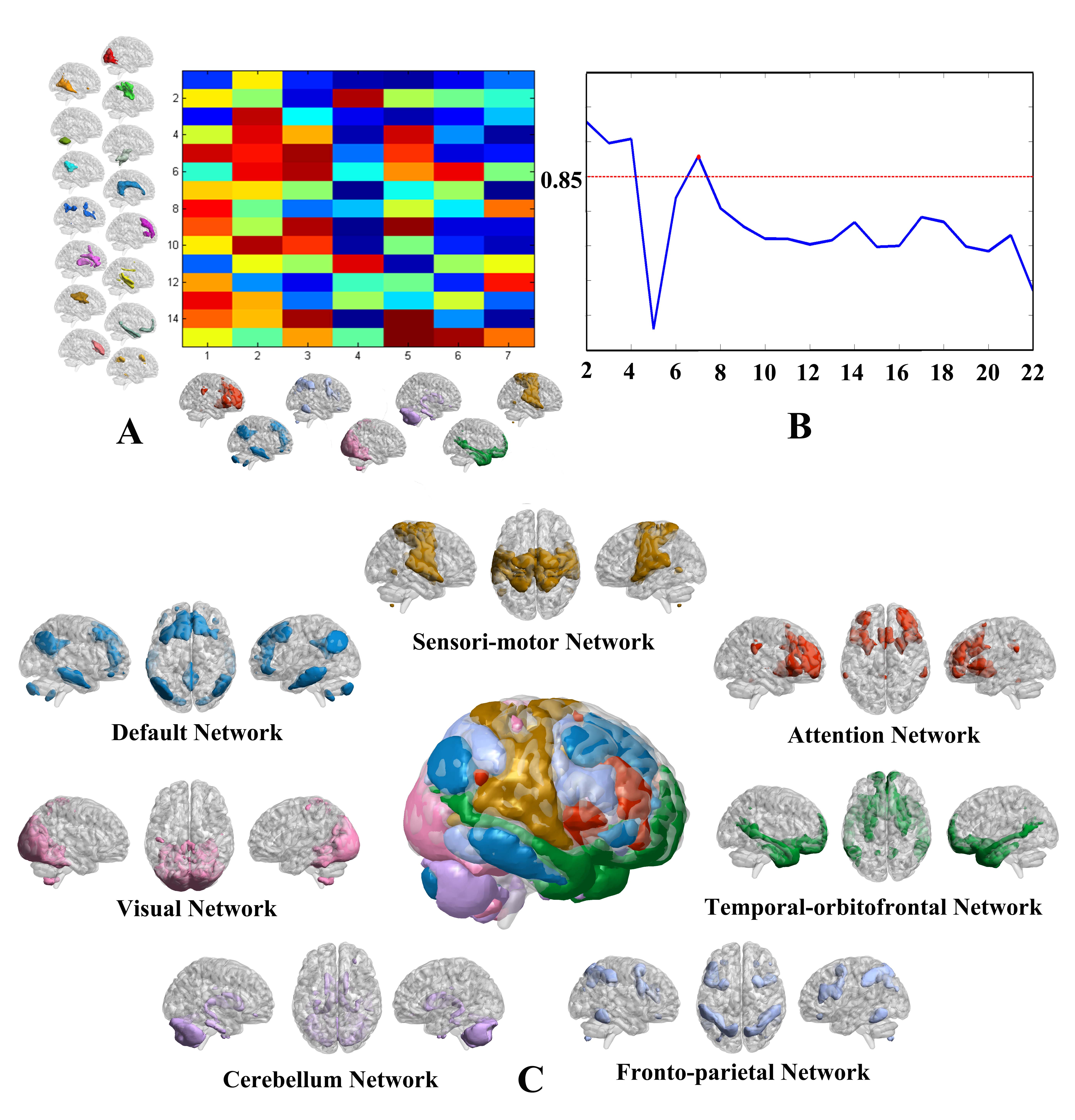
Resting-state functional Magnetic Resonance Imaging (RS-fMRI) was performed in 23 participants within 24 hours of flying from Hawaii, USA to Chengdu, China and again 50-days later. Gray (GM) and white matter (WM) networks were identified by K-means clustering [8] and three analyses were performed. WM functional connectivity (FC) and WM Functional Covariance Connectivity (FCC) [9, 10] analyzed. Next, a sliding window method was used to establish dynamic WM FC. WM static and dynamic FC and FCC were compared between when participants had initially completed their journey and 50-days later. Emotion was assessed by using the Positive and Negative Affect Schedule [11] and the State Anxiety Inventory [12].Results
Analysis using the Colombia Jet Lag Questionnaire confirmed that all 23 participants experienced Jet Lag and for which the most prominent symptom was drowsiness and anxious. A comprehensive analysis of the static and dynamic FC of WM resting state networks revealed the cingulate network (WM7) and the precentral/postcentral network (WM3) to be the brain networks most implicated in producing the symptoms associated with Jet Lag. In particular, after the long haul flight which produced Jet Lag analysis of the brain WM resting state networks reveals there to have occurred a significant reduction (i) in FC between WM7 and other white matter networks namely WM1, WM2, WM3, WM5, WM12 and WM13, and between WM3 and WM4, (ii) in FC between WM7 and GM4, (iii) in FCC between WM7 and WM3, (iv) in dynamic FC between WM7 and WM1, WM2, WM3 and WM12 for state 1 (as well as similar effects for states 2, 4 and 5), and (v) a significant negative correlation between positive emotion and FC between WM7 and WM3 and a highly significant correlation between positive emotion and FC between WM7 and GM7, and one will note that effects relating to WM7 and WM3 are present in all of (i) to (v). Additionally, the ventral frontal network (WM14) is implicated in that (i) there is a significant reduction in FC and between WM14 and GM4 and (ii) in dynamic FC between WM5 and WM10 and WM14 for state 1. In all cases FC decreases during Jet lag compared to recovery.Discussion
The cingulate network is known to play an important role in information processing [13] and a reduction in the FC of the cingulate network, especially with the precentral/postcentral network. The function of the cingulate has been reported to be disrupted in subjects with both sleep disorders [14] and mood disorders [13, 15]. Furthermore, a DTI study of participants categorized as either early (EC), late (LC) or intermediate (IC) chronotypes, where EC’s tend to wake up early in the morning and find it difficult to remain awake beyond their usual bedtime, and LC’s go to bed late and have difficulties getting up, revealed decreased white matter fractional anisotropy of cingulate gyrus in subjects with the LC chronotype [16] who have symptoms such as sleep disturbance and vulnerability to depression that are also associated with Jet Lag [16]. On the contrary subjects with sleep deprivation were reported to show increased FC [17] and increased FC density [35] in pre- and post-central regions bilaterally and which was suggested to reflect a compensatory mechanism [18]. At the time of experiencing Jet Lag participants showed significantly decreased positive emotion and increased anxiety, and with the former being negatively correlated with the FC of WM7 with both WM3 and GM7, suggesting that the cingulate may play a crucial role in expression of emotions and maintenance of positive mood.Conclusion
The results of this study provide further evidence for the existence of WM networks and extends the results of a previous study to show that Jet Lag is associated with alterations in static and dynamic WM FC and WM FCC especially in sensori-motor networks. Jet Lag is a complex problem which not only related with sleep rhythm but also influence emotion.Acknowledgements
This study was supported by the National Natural Science Foundation (Grant Nos. 81971595, 81771812, 81761128023 and 81621003). Program for Changjiang Scholars and Innovative Research Team in University (PCSIRT, Grant No. IRT16R52) of China, and the Science and Technology Department of Sichuan Province (2018SZ0391) and the Innovation Spark Project of Sichuan University (No. 2019SCUH0003).References
1. Westchester, I., The international classification of sleep disorders: diagnostic & coding manual (2nd ed). . American Academy of Sleep Medicine, 2005.
2. Morgenthaler, T.I., et al., Practice parameters for the clinical evaluation and treatment of circadian rhythm sleep disorders. An American Academy of Sleep Medicine report. Sleep, 2007. 30(11): p. 1445-59.
3. Sack, R.L., Clinical practice. Jet lag. N Engl J Med, 2010. 362(5): p. 440-7.
4. Drust, B., et al., Circadian rhythms in sports performance--an update. Chronobiol Int, 2005. 22(1): p. 21-44.
5. Drust B, W.J., Atkinson G, Edwards B, Reilly T., Circadian rhythms in sports performance--an update. Chronobiol Int. , 2005. 22(1): p. 21-44.
6. Herxheimer, A., Jet lag. BMJ Clin Evid, 2014.
7. Gander PH, N.D., Rosekind MR, Connell LJ., Age, circadian rhythms, and sleep loss in flight crews. Aviat Space Environ Med. , 1993. 64(3): p. 189-195.
8. Peer, M., et al., Evidence for Functional Networks within the Human Brain's White Matter. The Journal of Neuroscience, 2017. 37(27): p. 6394-6407.
9. Jiang, Y., et al., Dysfunctional white-matter networks in medicated and unmedicated benign epilepsy with centrotemporal spikes. Hum Brain Mapp, 2019.
10. Zhang, H., et al., Topographical Information-Based High-Order Functional Connectivity and Its Application in Abnormality Detection for Mild Cognitive Impairment. J Alzheimers Dis, 2016. 54(3): p. 1095-1112.
11. Crawford, J.R. and J.D. Henry, The positive and negative affect schedule (PANAS): construct validity, measurement properties and normative data in a large non-clinical sample. Br J Clin Psychol, 2004. 43(Pt 3): p. 245-65.
12. Ramanaiah, N.V., M. Franzen, and T. Schill, A psychometric study of the State-Trait Anxiety Inventory. J Pers Assess, 1983. 47(5): p. 531-5.
13. Amir, N., et al., Increased activation of the anterior cingulate cortex during processing of disgust faces in individuals with social phobia. Biol Psychiatry, 2005. 57(9): p. 975-81.
14. Tomasi, D., et al., Impairment of attentional networks after 1 night of sleep deprivation. Cereb Cortex, 2009. 19(1): p. 233-40.
15. Wang, T., et al., Increased insular connectivity with emotional regions in primary insomnia patients: a resting-state fMRI study. Eur Radiol, 2017. 27(9): p. 3703-3709.
16. Rosenberg, J., et al., "Early to bed, early to rise": diffusion tensor imaging identifies chronotype-specificity. Neuroimage, 2014. 84: p. 428-34.
17. Liu, C.H., et al., Increased Posterior Insula-Sensorimotor Connectivity Is Associated with Cognitive Function in Healthy Participants with Sleep Complaints. Front Hum Neurosci, 2018. 12: p. 35.
18. Huang, Z., et al., Abnormal amygdala connectivity in patients with primary insomnia: evidence from resting state fMRI. Eur J Radiol, 2012. 81(6): p. 1288-95.
Figures
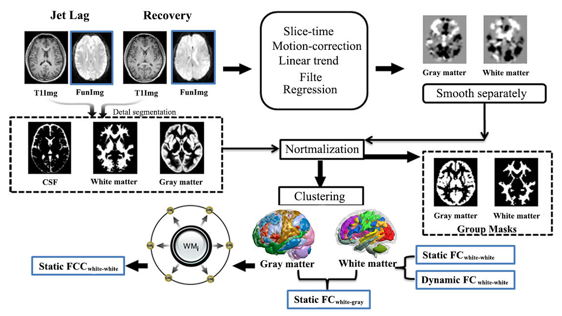
Flow chart of consecutive steps in the processing of the RS-fMRI data. The RS-fMRI images were segmented into WM and GM by using information of from the 3D T1-weighted image and K-means clustering analysis was used to identify WM and GM networks. Finally, individual static WM FC, dynamic WM FC and FCC, as well as FC between WM and GM networks were measured.
GM, gray matter; WM, white matter; FCC, functional covariance connectivity; FC, functional connectivity