2037
Effect of memory retrieval-extinction procedure on the brain of patients in smoking cessation revealed by resting-state fMRI1Department of Medical Imaging and Radiological Sciences, Chang Gung University, Taoyuan, Taiwan, 2Department of Psychology, Chung Shan Medical University, Taichung, Taiwan, 3Clinical Psychological Room, Chung Shan Medical University Hospital, Taichung, Taiwan, 4Medical Imaging Research Center, Institute for Radiological Research, Chang Gung University and Chang Gung Memorial Hospital at Linkou, Taoyuan, Taiwan, 5Department of Psychiatry, Chang Gung Memorial Hospital, Chiayi, Taiwan
Synopsis
Previous studies used memory retrieval-extinction procedure to successfully reduce the drug self-administration behavior in rats and heroin patients. The method was rarely used in smoking cessation. This study used resting-state fMRI to evaluate the effect of memory retrieval-extinction procedure on heavy smokers. We found that lower default mode network activity and higher reward system activation in smokers who finished the memory retrieval-extinction procedure. It may reflect a nonpharmacological method induced the smoking-related emotions but reduced smoking memory retrieval. In conclusion, our results could give a new perspective on clinical treatment for smoking cessation.
Introduction
Cigarette smoking causes many serious health problems and premature death. According to the report of the World Health Organization in 2018, tobacco kills more than 8 million people each year around the word1. Nicotine replacement therapy (NRT) is one of the most common methods to treat for smoking cessation. However, most smokers could eventually not quit smoking. We aimed to evaluate whether the memory retrieval-extinction procedure as an adjunct treatment reduced nicotine cravings and increased motivation to quit smoking.Methods
Participants and experimental designSixty-seven heavy smokers were recruited from the smoking cessation outpatient service of National Cheng Kung University Hospital. 32 of them were randomly assigned to receive the memory retrieval-extinction procedure (SRE), and the remaining 35 were not (SN). Under the same inclusion and exclusion criteria, 43 non-smokers (HC) were recruited as healthy controls.
For the memory retrieval-extinction manipulation, smokers watched the videos for 5 minutes to retrieve the smoking memories, and then 10 min delayed before the 45 minutes cues of the extinction sessions. The cue for SRE was smoking-related pictures, and the cue for SN was landscape pictures.
Image acquisition
All participants were conducted on a 3-Tesla MRI system (GE Medical Systems, Waukesha, WI, USA) to obtain the Blood Oxygenation Level Dependent (BOLD) signals. The resting-state functional images were acquired using a T2* weighted echo planar image (EPI) sequence with the following parameter: TR/TE = 2000 ms / 30 ms, FOV = 224 mm 224 mm2, matrix size = 64 64, slice thickness = 3.5 mm.
Image processing and analysis
Processing was performed using Statistical Parametric Mapping 12 (SPM12, Wellcome Department of Cognitive Neurology, Landon, UK). Image processing was performed by following steps: 1) slice-timing correction was used for slightly different image timing for each slice acquisition; 2) realignment were performed for the head motion correction; 3) normalization were used with the ICBM- East Asian Brain space template and affine regularization; and 4) images were smoothed by Gaussian kernel (FWHM: 7mm) to compensate for differences in normalization between individuals and increase the signal-to-noise ratio.
The band-pass filter with frequency from 0.01 to 0.12 Hz was applied to remove physiological noise by using Resting-State fMRI Analysis toolkit (REST v1.8, http://www.restfmri.net). A regional homogeneity (ReHo) method was then used to calculate locally spontaneous synchronization of brain activity and the amplitude of low-frequency fluctuation (ALFF) approach was measured by the spontaneous neural activity in low-frequency to possess physiological significance. To evaluate group differences in mReHo and mfALFF, we used two-sample t-tests in SPM12. Besides, the multiple regression was performed between brain activity and questionnaires, including the Fagerström Test for Nicotine Dependence (FTND) and the Drug Use Disorders Identification Test- Extended (DUDIT-E).
We also explored the brain networks with graph theory by using the functional connectivity toolbox (CONN, https://web.conn-toolbox.org). We used an automated anatomical labeling (AAL) template to define the network nodes and divided the brain into 45 regions per hemisphere as ROIs. The graph analysis toolbox (GAT, https://www.nitrc.org/projects/gat) was then performed to analyze the topological properties of the functional brain network.
Results
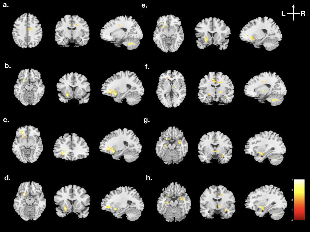
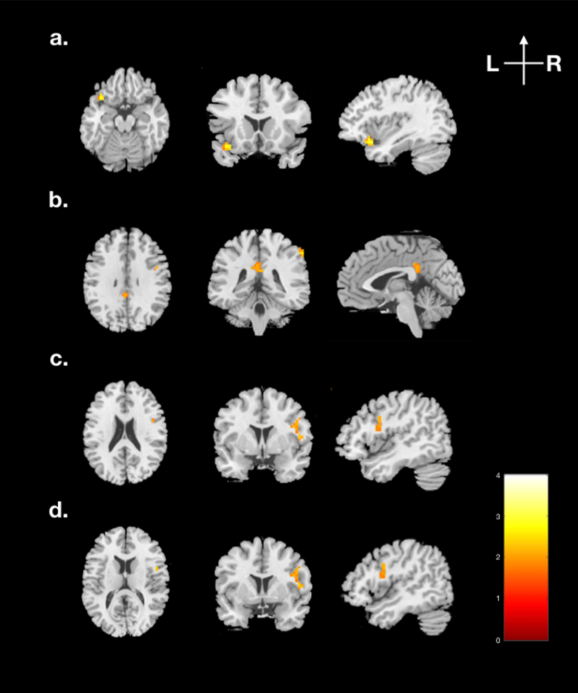
Group comparison between the SRE and SN in mReHoAs shown in Fig. 1, the SRE showed significantly higher activation in the bilateral midcingulate cortex (MCC), left superior frontal gyrus (SFG), left inferior frontal gyrus (IFG), left insula, left putamen, right thalamus, right amygdala, and bilateral hippocampus than SN with p-value < 0.01. In Fig. 2, the SRE showed significantly lower activation than SN in the left superior temporal gyrus (STG), left posterior cingulate cortex (PCC), right IFG and right rolandic operculum with p-value < 0.02.
Group comparison between the SRE and SN in mfALFF
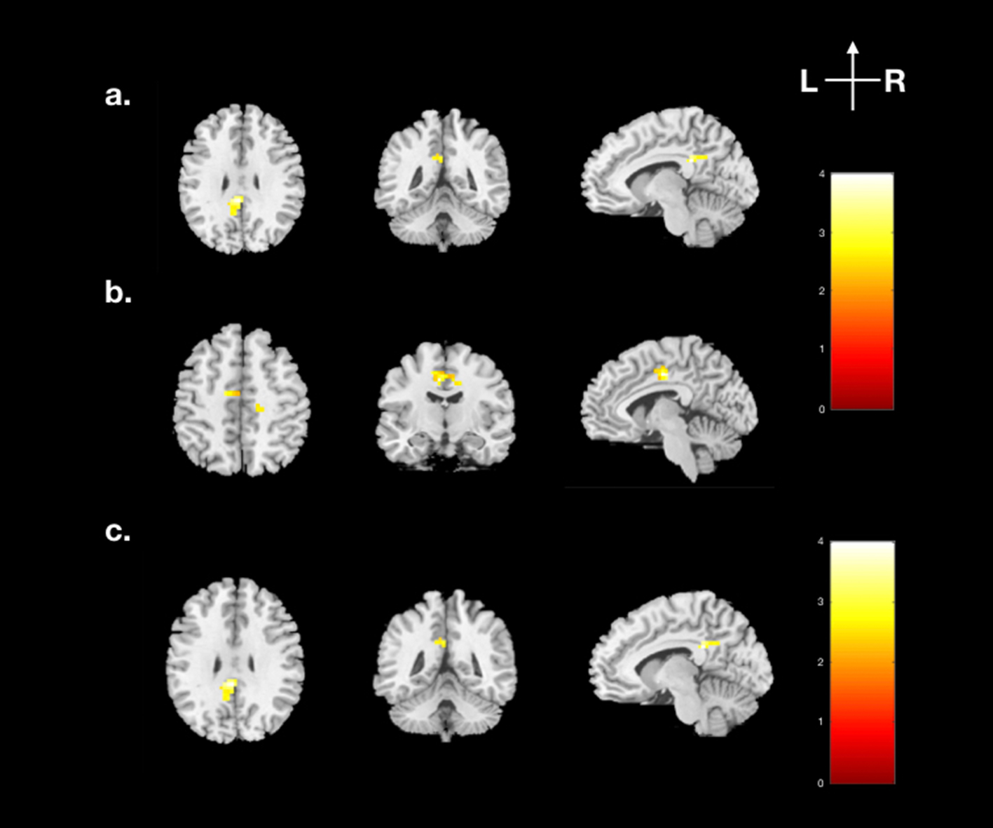
As shown in Fig. 3, the significantly higher activation in the left supplementary motor area (SMA) and bilateral MCC with p-value < 0.01 were found in SRE. The SRE also showed significantly lower activation in the left PCC with p-value < 0.01.
Graph theoretical analysis between smokers and HC
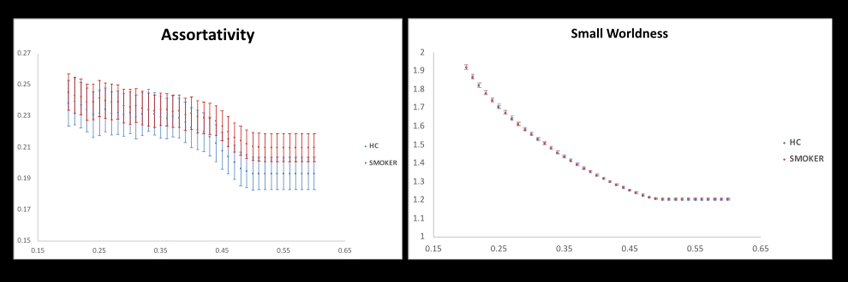
As shown in Fig. 4, we found a significant larger assortativity in smokers compared with HC. It may probably indicate the functional brain network is more flexibility under brain damages2.
Discussion
The previous studies showed the higher activation in IFG, SMA, and putamen was associated with the links between cravings and subsequent smoking were attenuation3. Our results confirmed that higher activation in the frontal cortex, nucleus accumbens, cingulate and amygdala was involved in reward processing and nicotine present4. The parts of default mode network (DMN) regions in our result showed lower activation during the SRE, which was consistent with previous studies that DMN deactivated during the cognitive tasks5, and the PCC involved in memory retrieval and craving6.Conclusion
The present study is one of few explore the memory retrieval-extinction procedure in smoking cessation. Our fMRI results provide evidence to support the memory retrieval-extinction procedure effectively induced the smoking-related emotions but reduced the smoking memory retrieval to clinical treatment with important insight for smoking cessation.Acknowledgements
This study was supported by the research program MOST105-2410-H-040-001-MY3, which were sponsored by the Ministry of Science and Technology, Taipei, Taiwan.References
1. World Health Organization.WHO Report on the Global Tobacco Epidemic, 2019. Geneva: World Health Organization; 2019.
2. Chang-hyun Park, Soo Yong Kim, Yun-Hee Kim, et al. Comparison of the small-world topology between anatomical and functional connectivity in the human brain. Physica A. 2008; 387: 5958–5962.
3. Elliot T. Berkman, Emily B. Falk, Matthew D. Lieberman. In the Trenches of Real-World Self-Control: Neural Correlates of Breaking the Link Between Craving and Smoking. Psychological Science. 2011; 22(4): 498–506.
4. Elliot A. Stein, John Pankiewicz, Harold H. Harsch, et al. Nicotine-Induced Limbic Cortical Activation in the Human Brain: A Functional MRI Study. Am J Psychiatry. 1998; 155(8): 1009–1015.
5. Michael D. Fox, Abraham Z. Snyder, Justin L. Vincent, et al. The human brain is intrinsically organized into dynamic, anticorrelated functional networks. PNAS. 2005; 102(27): 9673–9678.
6. Keliane Liberman, Peter Van Schuerbeek, Sarah Herremans, et al. The effect of nicotine patches on craving in the brain: A functional MRI study on heavy smokers. Medicine. 2018; 97: 39.
Figures