2024
Application of Deep Learning Reconstruction to Compressed-sensing Thin-slice Fat-suppressed T2-weighted Imaging of the Orbit1Department of Diagnostic Imaging and Nuclear Medicine, Kyoto University Graduate School of Medicine, Kyoto, Japan, 2MRI Systems Division, Canon Medical Systems Corporation, Otawara, Japan, 3Human Brain Research Center, Kyoto University Graduate School of Medicine, Kyoto, Japan, 4Department of Advanced Medical Imaging Research, Kyoto University Graduate School of Medicine, Kyoto, Japan
Synopsis
Deep learning reconstruction (DLR) is a novel denoising processing. We applied DLR to a compressed sensing (CS) sequence of orbital thin-slice fat-suppressed T2-weighted imaging with one number of excitation (NEX). A CS sequence with one NEX without DLR and a conventional sequence with two NEX were also obtained to evaluate the denoising performance. Combined usage of DLR with CS reduced image noise and improved the image quality of the optic nerves and the medial rectus muscles, while achieving shorter acquisition time, compared with the CS and the conventional sequences without DLR.
Introduction
Orbital fat-suppressed T2-weighted imaging is an essential MR sequence for evaluating patients with optic neuritis 1 and thyroid-associated ophthalmopathy.2 The role of MR imaging is to detect high signal intensity as well as swelling of the optic nerves (ON) and the extraocular muscles. However, MR imaging of the orbit is susceptible to eye movement artifact during the scan. Compressed sensing (CS) enables accelerated MR acquisition through a pseudo-random undersampling of k-space.3 Artifacts due to the undersampling are removed by a sophisticated reconstruction algorithm.4 However, too much undersampling may lead to low signal-to-noise ratio (SNR). It has recently been reported that deep learning reconstruction (DLR) can reduce image noise using a different strategy from CS.5 By combining CS and DLR together, we could achieve faster image acquisition without increasing the image noise. In this study, we applied DLR to a CS sequence of orbital thin-slice fat-suppressed T2-weighted imaging with one number of excitation (NEX). A CS sequence with one NEX without DLR (CS-NEX1) and a conventional sequence with two NEX (NEX2) were also obtained to evaluate the denoising performance. The image quality of ON and the medial rectus muscles (MRM) was evaluated.Methods
This prospective observation study was approved by our Institutional Review Board, and written informed consent was obtained.Subjects
Twelve adults (6 males and 6 females; mean age, 72; range, 53–84 years) were enrolled who underwent MR examination of the orbit.
Image acquisition and reconstruction
MR imaging was obtained using a 3T unit (Vantage Galan 3T / ZGO, Canon Medical Systems Corporation, Otawara, Japan) equipped with a 32-channel head coil. We acquired coronal fast spin-echo fat-suppressed T2-weighted imaging with CS-NEX1 and NEX2. The acquisition parameters were as follows: repetition time, 4219 ms for CS-NEX1 and 4505 ms for NEX2; echo time, 60 ms for CS-NEX1 and 77 ms for NEX2; flip angle, 89°; slice thickness, 2 mm; slice gap, 1 mm; field of view, 10 × 10 cm; matrix, 320 × 320; in-plane resolution, 0.31 × 0.31 mm; bandwidth, 139.5 Hz/pixel for CS-NEX1 and 162.7 Hz/pixel for NEX2; 11 slices; and scan time, 1 m 29 s for CS-NEX1 (acceleration factor of CS = 2) and 4 m 35 s for NEX2. CS-NEX1 was further processed with DLR, which yielded DLR-CS-NEX1.
Image analysis
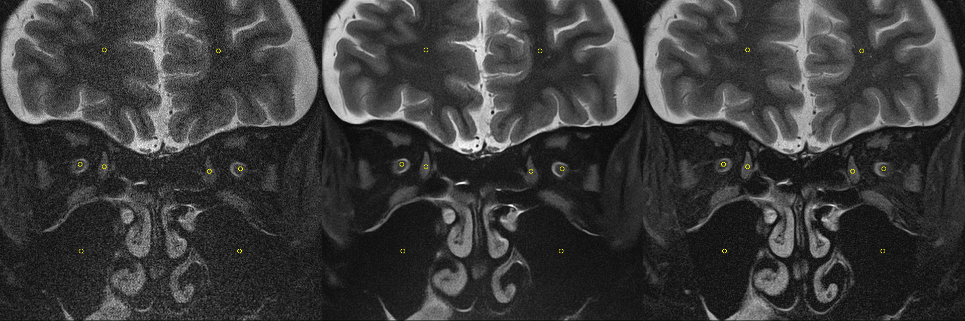
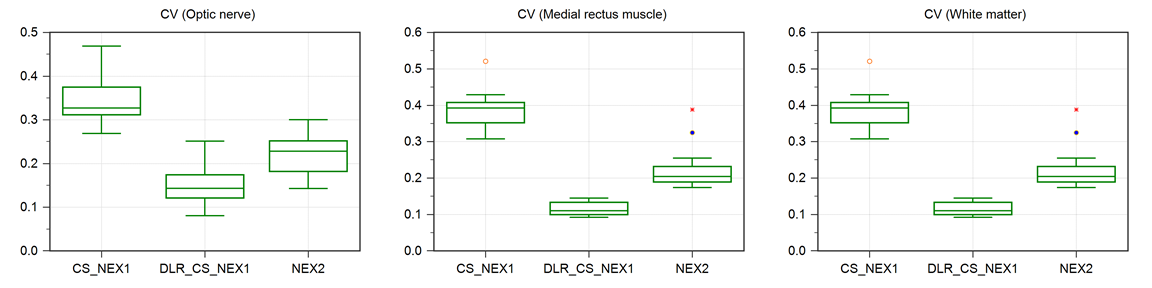
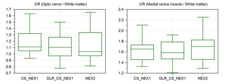
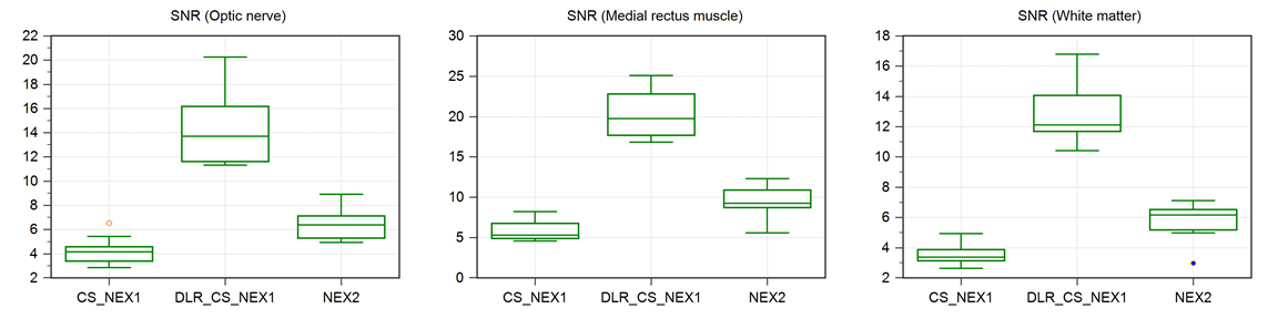
All images were analyzed using ImageJ (https://imagej.nih.gov/ij/). To examine quantitatively, regions of interest (ROIs) were manually placed on ON, MRM, white matter (WM) and paranasal sinuses in a representative slice (Figure 1). The degree of homogeneity in ON, MRM and WM were evaluated by the coefficient of variation (CV, the standard deviation divided by the mean). The mean ROI values of ON and MRM were divided by that of WM, which yielded contrast ratios (CRs) of ON/WM and MRM/WM, respectively. For the tissue SNR, the mean ROI values of ON, MRM and WM were divided by the standard deviation of paranasal sinuses, which is assumed to contain only the background noise. For qualitative assessment, a neuroradiologist with an experience of 14 years rated CS-NEX1, DLR-CS-NEX1 and NEX2 using a four-point scale (1 = poor, 2 = fair, 3 = good, 4 = excellent).
Statistical analysis
The differences among CS-NEX1, DLR-CS-NEX1 and NEX2 were compared statistically using a Friedman test. A p-value of 0.05 was considered to indicate a presence of statistical significance.
Results
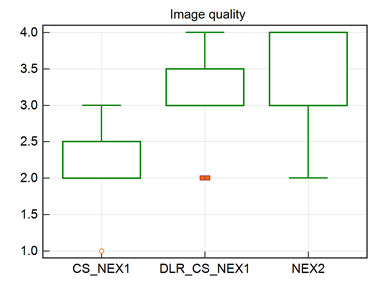
DLR-CS-NEX1 demonstrated significantly lower CV than NEX2, and NEX2 showed significantly lower CV than CS-NEX1 (Figure 2). No significant difference was found among the three images in terms of CR (Figure 3). DLR-CS-NEX1 demonstrated significantly higher SNR than NEX2, and NEX2 showed significantly higher SNR than CS-NEX1 (Figure 4). DLR-CS-NEX1 and NEX2 demonstrated better visual image quality than CS-NEX1, but no significant difference was found between DLR-CS-NEX1 and NEX2 in terms of visual image quality (Figure 5).Discussion
High-resolution T2-weighted imaging with a thin slice thickness of 2 mm is clinically useful for evaluation of the optic nerves and the extraocular muscles since partial volume effect will be minimized. In clinical practice, patients are instructed to see the target point so as not to move eyes, however, a certain motion artifact associated with eye movement is inevitable in MR images with longer acquisition time. High-resolution MR imaging with shorter scan time will be beneficial for patient care. This study demonstrated that DLR could further reduce image noise left in CS reconstruction and improve the image quality of the orbit. Theoretically, acceleration by parallel imaging combined with CS may cause SNR reduction compared with full sampling. DLR could play a complimentary role with CS. A limitation existed, however, as patients with optic neuritis or orbital diseases were not included in this preliminary study. Further clinical studies are required for evaluation of DLR application to high-resolution T2-weighted imaging.Conclusion
Combined usage of DLR with CS reduced image noise and improved the image quality of the orbit on thin-slice fat-suppressed T2-weighted imaging, while achieving acquisition time, compared with CS and conventional sequences without DLR.Acknowledgements
No acknowledgement found.References
1. Jackson A, Sheppard S, Laitt RD, et al. Optic neuritis: MR imaging with combined fat- and water-suppression techniques. Radiology 1998;206:57-63
2. Lo C, Ugradar S, Rootman D. Management of graves myopathy: Orbital imaging in thyroid-related orbitopathy. J AAPOS 2018;22:256.e251-256.e259
3. Jaspan ON, Fleysher R, Lipton ML. Compressed sensing MRI: a review of the clinical literature. Br J Radiol 2015;88:20150487
4. Lustig M, Donoho D, Pauly JM. Sparse MRI: The application of compressed sensing for rapid MR imaging. Magn Reson Med 2007;58:1182-1195
5. Kidoh M, Shinoda K, Kitajima M, et
al. Deep Learning Based Noise Reduction for Brain MR Imaging: Tests on Phantoms
and Healthy Volunteers. Magn Reson Med
Sci 2019
Figures