2022
Small-World networks in the human brain revealed by cortical layers from T1 mapping1MR Research, GE Healthcare, Beijing, China, 21Department of Biomedical Engineering, Peking University, Beijing, China, 3School of Data and Computer Science, Sun Yat-Sen University, Guangzhou, China, 4Department of Clinical Medicine, Faculty of Medicine and Health Sciences, Macquarie University, Sydney, Australia, 5Department of Medical Imaging, Sun Yat-sen University Cancer Center, Guangzhou, China
Synopsis
Our goal is to propose a new approach to construct the brain anatomical connectivity networks using T1 mapping, and verify whether this novel brain network has higher Small-Worldness and efficiency organization. We reviewed the previous studies, and discovered that the cortical layers could be delineated on the basis of myeloarchitecture, while T1 mapping strongly correlated with intro-cortical myelin contents. Hence, it is feasible to quantify the anatomical connectivity using T1 mapping. Our resulted supported our hypothesis and highlighted the higher Small-Worldness and efficiency of this network compared with conventional macroscale cortical morphometry based approach.
Introduction
Analysis of human connectome using MRI-based morphometric metrics, such as cortical thickness and volume, have gained much attention in recent years. Since this work first proposed by He et al. in 2007,1 it has been rapidly applied to discover the neural underpinnings of human cognition and neurological disorders. However, the mesoscale characterization of cortical morphometry based on T1W MRI is hard to investigate, such as the cortical layers, columns, stripes. Previous study indicated that the six-layer organizations of the whole neocortex could not only be delineated on the basis of cytoarchitecture, but also myeloarchitecture, which suggested that myelin properties may also be cortical-layer dependent.2 In addition, research scientists suggested that T1 mapping may be a good choice to discover the six cortical layers in the mesoscale.3 Furthermore, a direct evidence comes from recent study indicates that similarity of areas according to intracortical myelin content is a good predictor for whether two areas are functionally connected.4 Hence, we have a hypothesis that the small-world architectures of the cortical anatomical connectivity based on T1 mapping is more efficient than conventional macroscale cortical measures. To test this hypothesis, synthetic MRI with simultaneously generating T1, T2 and PD maps were applied to 21 healthy participants in current study.Methods
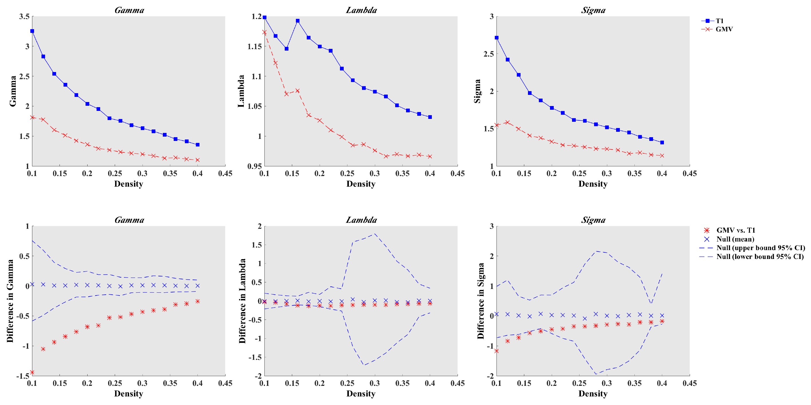
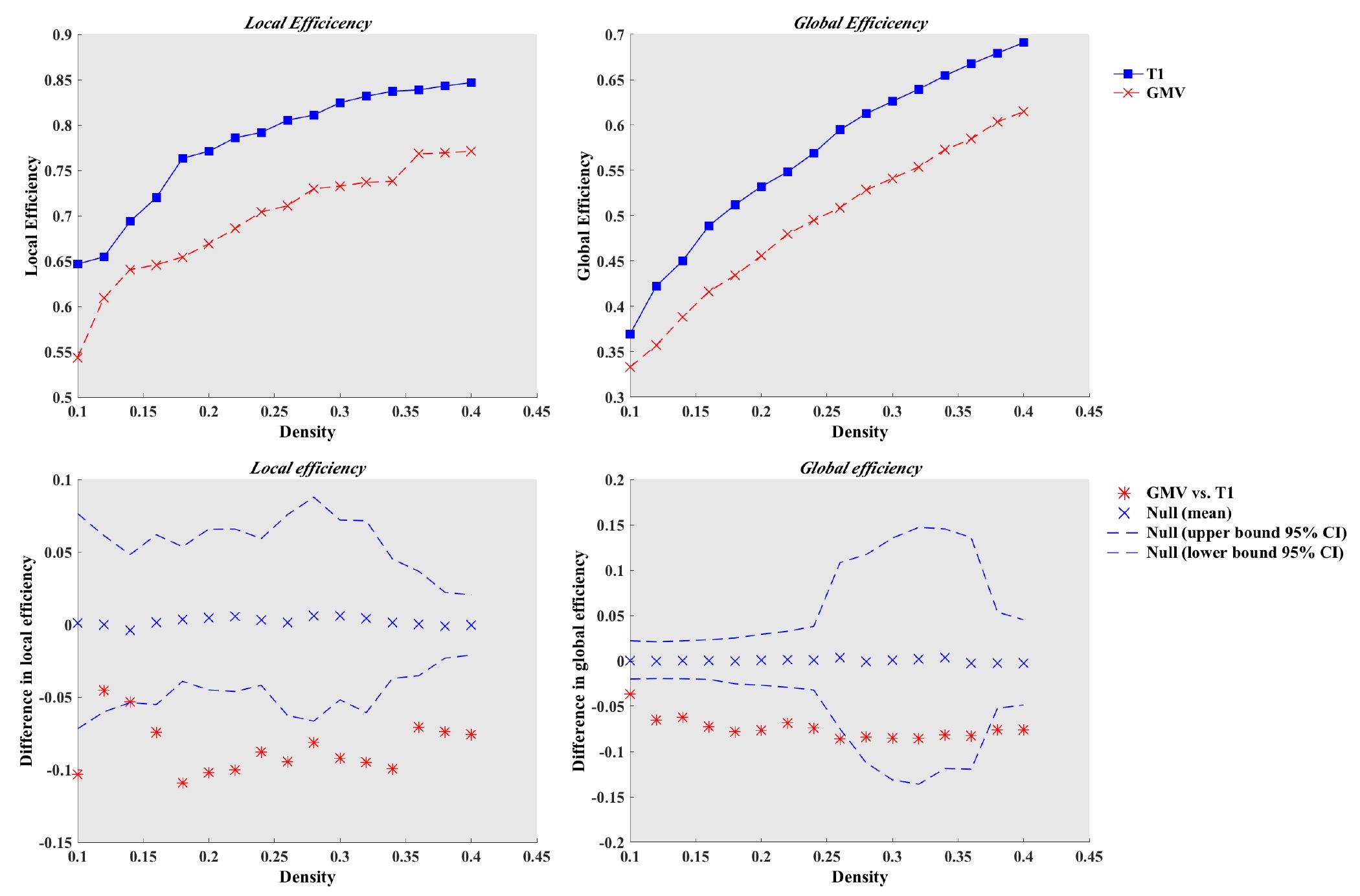
For each subject, MR scans were performed on a 3.0T whole body scanner (Signa Pioneer, GE, WI) with a 32-channel head coil. Axial images were acquired using 3D BRAVO sequence and synthetic MRI with spatial resolution equal to 1mm and 2mm isotropic, respectively. After MRI scanning, the relaxation quantitative maps were generated using synthetic MRI software. The T1 images were analyzed with VBM8 toolbox, then the cortical volumes were calculated by masking the 90 former ROIs of AAL atlas with the individually modulated and normalized GM images. To extract the atlas based quantitative values, T1 anatomical images of each subjects were first co-registered to T1 map. Then, the co-registered T1 images were normalized to MNI space using SPM12. Thereafter, T1 map could be transformed to MNI space. Last, the AAL atlas were applied to normalized T1 image to extract the values of the former 90 regions. With regard to the network analysis, it is known that two key metrics were applied to described the complex networks in the human brain: clustering coefficient (CP) and characteristics path length (LP). To evaluate the small-world properties, CP and LP of the real network were compared with corresponding random networks (CPrand and LPrand). Typically, a small-world organization should fulfill the following conditions: Gamma = CPreal/CPrand > 1 and Lambda = LPreal/LPrand ~ 1. For convenience, a simple quantitative measurement, Small-Worldness, Sigma = Gamma/Lambda > 1, was applied to represent the proxy of small-world properties. In addition, to compare the effectiveness of information transfer, the Global and Local efficiency were also calculated. The network construction and analysis of both the cortical volumes and cortical layers based methods were using Graph Analysis Tools (GAT). To compare the differences of topological measures between the two networks, a nonparametric permutation test with 1000 repetitions was applied. A value of P < 0.05 were considered statistically significant.Results and discussion
Our results showed that both the cortical volumes and cortical layers based approach had small-world architectures (Figure 1). However, the Small-Worldness index of cortical layers connectivity networks is significant higher than cortical volumes based network (Figure 1). Our results provided us evidences that the cortical layers based anatomical connectivity networks had greater local interconnectivity and short mean distance between regions. In addition, both the global and local efficiency showed significant increased values in the networks constructed using T1 mapping compared with cortical volumes (Figure 2). Network efficiency quantifies the effectiveness of information transfer within brain network. We know that human brain is an economic system, the higher global and local efficiency were more in line with this intrinsic architecture. Hence, we speculated that the anatomical associations based on intra-cortical myelin content may reflect the actual mechanism of brain information transmission more sensitively than macroscale cortical morphometry. Our results were also supported by previous study, which suggests that cortical areas with similar microstructure are more likely to be connected.Conclusion
Our research first proposed a novel anatomical connectivity network with small-world organization using cortical layers from T1 mapping. Importantly, we highlighted the higher Small-Worldness and efficiency of this network compared with conventional macroscale cortical morphometry based approach.Acknowledgements
No acknowledgement found.References
1. He Y, Chen ZJ, Evans AC. Small-world anatomical networks in the human brain revealed by cortical thickness from MRI. Cerebral cortex. 2007; 17(10): 2407-19.
2. Micheva KD, Wolman D, Mensh BD, et al. A large fraction of neocortical myelin ensheathes axons of local inhibitory neurons. elife. 2016; 5: e15784.
3. Lifshits S, Tomer O, Shamir I, et al. Resolution considerations in imaging of the cortical layers. NeuroImage. 2018; 164: 112-20.
4. Huntenburg JM, Bazin PL, Goulas A, et al. A systematic relationship between functional connectivity and intracortical myelin in the human cerebral cortex. Cerebral Cortex. 2017; 27(2): 981-97.
Figures