2005
Micro-Structural Delineation of the Human Olfactory Bulb with High Field MR Microscopy.1Department of Anatomy and Medical Imaging, University of Auckland, Auckland, New Zealand, 2Laboratory of Functional and Molecular Imaging, NINDS, National Institutes of Health, Bethesda, MD, United States
Synopsis
Measuring anatomical structures in the olfactory bulb may be useful in studying neurodegenerative diseases such as Alzheimer’s and Parkinson’s disease. Preliminary magnetic resonance microscopy images of the olfactory bulb and tract have been obtained at high resolution (19-25 μm isotropic) and shown to have features that correlate well with immunohistochemical staining.
Introduction
In neurodegenerative diseases such as Alzheimer’s and Parkinson’s disease, olfactory deficits preceed the characteristic symptoms such as memory loss and tremor by many years. The sensory neurons that detect smell are located in the roof of the nose and project directly to the olfactory bulb in the brain. Therefore, it is hypothesized that environmental insults that may contribute to neurodegenerative diseases may access the brain through the olfactory system. Furthermore, recent studies have shown that beta-amyloid, tau and alpha synuclein aggregates (the abnormal proteins in Alzheimer’s and Parkinson’s disease) accumulate in the olfactory bulb in these diseases. Despite this evidence for an olfactory component to neurodegenerative disease pathology, alterations to the structure of the human olfactory bulb in these diseases have not yet been determined. Very little work has studied the human olfactory bulb in a clinical setting in the context of ageing and neurodegeneration (1–4). Due to their small size and location underneath the brain, the olfactory bulbs can be difficult to visualize using standard clinical MRI. Furthermore, good quality post-mortem specimens are rare due to their poor structural integrity and tendency to be ripped or distorted as the brain is removed from the skull. As such we are aware of only one study of post-mortem human olfactory bulbs which showed that high-field MRI (9.4T) can be used to show some layer contrast in the bulb, although this study acquired these scans at relatively low resolution (100 μm) (5). Here we present preliminary scans on formalin-fixed human olfactory bulbs using MR microscopy (MRM) that show higher resolution imaging can be used to identify several structural features within the bulb.Methods
Pairs of olfactory bulbs were obtained from the Neurological Foundation of New Zealand Human Brain Bank at the University of Auckland, Centre for Brain Research. Bulbs were moved from 5% to 1% formalin and soaked in 50 μM MnCl2 for 24 hours solution prior to MRM (6). For imaging, bulbs were embedded in agarose for stability. 3D gradient-echo images were acquired with single-echo at 19-25 μm isotropic or mutliple gradient echo at 25-30 μm isotropic. Images were acquired at either 11.7T or 14T. The images at 11.7T were acquired using a custom-built long solenoid allowing complete coverage of the bulb and the olfactory tract (~5 cm in length). At 14T, a 10-mm birdcage coil was used, covering around 2 cm of the bulb length. For the single-echo image TE was set at approximately the average T2* (between 10-15 ms) of the tissue based on a lower resolution multi-echo acquisition.Results
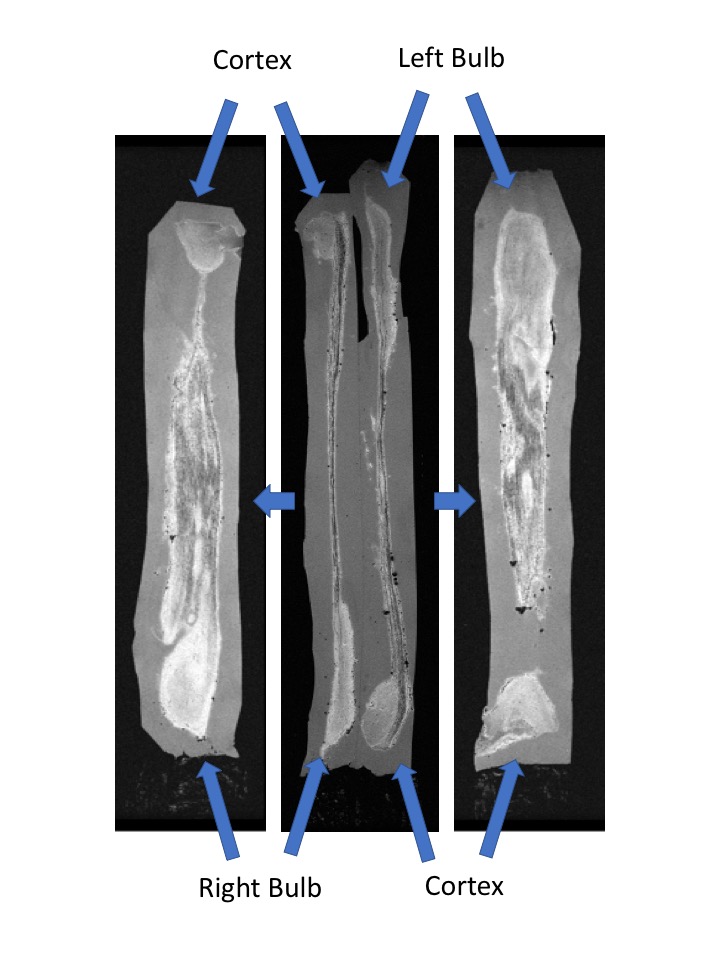
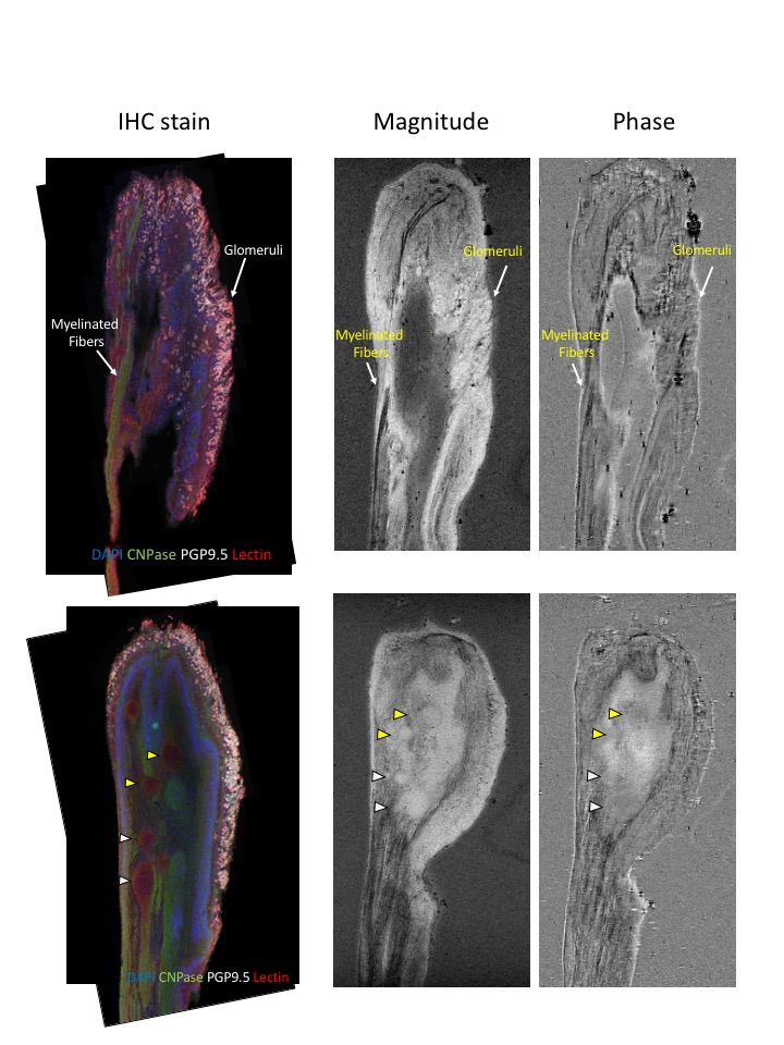
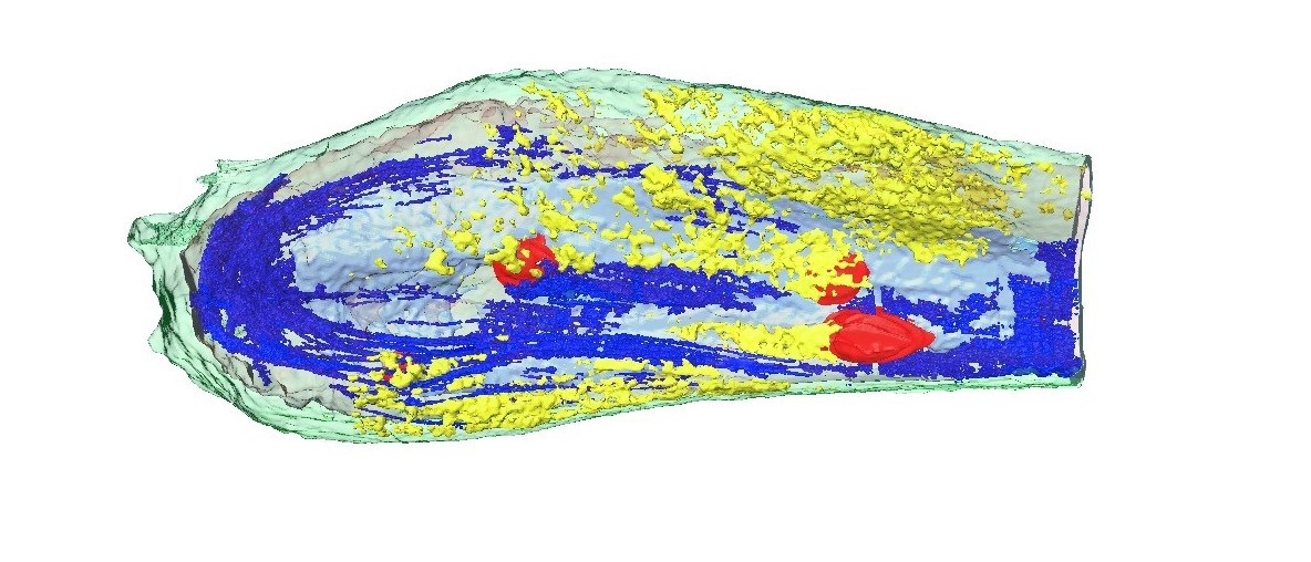
Figure 1 shows an example of the type of image obtainable, here at 20 μm isotropic resolution, with both the bulb and olfactory tract present for both left and right bulb from the same subject. White matter bundles are readily observed in the tract as well as the anterior olfactory nucleus (AON). (Imaging parameters: TR/TE = 30/12 ms, matrix size = 2560x384x384, FA = 15 degrees, scan time = 48 hrs). Figure 2 shows an image taken at 14T at 19 μm nominal resolution isotropic. (Imaging parameters: TR/TE = 30/10 ms, matrix size = 1280x512x320, FA = 15 degrees, scan time = 40 hrs). Structures visible include the anterior olfactory nucleus, white matter bundles, and laminar structure including the glomerular layer (and possibly individual glomeruli). Fluorescent immunohistochemistry was used to confirm the structures identified from the magnitude and phase images. Segmentation of the MR image in figure 2 was performed using AMIRA and the component tissues 3D surface rendered shown in Figure 3.Discussion
Magnetic resonance microscopy is well suited to study the olfactory bulb, as demonstrated here with basic image acquisition at high resolution. Currently, we are working toward quantitating our images with multiple echo datasets, so as to determine the origins of contrast and leading to improved clinical imaging of the bulb.Acknowledgements
No acknowledgement found.References
1. Segura B, Baggio HC, Solana E, Palacios EM, Vendrell P, Bargalló N, et al. Neuroanatomical correlates of olfactory loss in normal aged subjects. Behav Brain Res 2013; 246:148-53
2. Thomann PA, Santos V Dos, Seidl U, Toro P, Essig M, Schröder J, et al. MRI-Derived Atrophy of the Olfactory Bulb and Tract in Mild Cognitive Impairment and Alzheimer’s Disease. J Alzheimer’s Dis 2009; 17(1):213-21
3. Thomann PA, Santos V Dos, Toro P, Schönknecht P, Essig M, Schröder J. Reduced olfactory bulb and tract volume in early Alzheimer’s disease-A MRI study. Neurobiol Aging 2009;30(5):838-41
4. Servello A, Fioretti A, Gualdi G, Di Biasi C, Pittalis A, Sollaku S, et al. Olfactory Dysfunction, Olfactory Bulb Volume and Alzheimer’s Disease: Is There a Correlation? A Pilot Study. J Alzheimer’s Dis 2015;48(2):395-402
5. Burmeister HP, Bitter T, Heiler PM, Irintchev A, Fröber R, Dietzel M, et al. Imaging of lamination patterns of the adult human olfactory bulb and tract: In vitro comparison of standard- and high-resolution 3T MRI, and MR microscopy at 9.4T. Neuroimage 2012 Apr 15;60(3):1662-70
6. Manipulation of tissue contrast using contrast agents for enhanced MR microscopy in ex vivo mouse brain. Huang S, Liu C, Dai G, Kim YR, Rosen BR. Neuroimage. 2009 Jul 1;46(3):589-99
Figures