2002
Automatic segmentation of thalamic nuclei from Multi-Echo MP2RAGE acquisition at 7T1Children's Hospital of Philadelphia, Philadelphia, PA, United States, 2University of Pennsylvania, Philadelphia, PA, United States, 3University of Arizona, Tucson, AZ, United States
Synopsis
The efficacy of the Thalamus Optimized Multi-Atlas Segmentation (THOMAS) algorithm for segmentation of thalamic nuclei with multi-echo MP2RAGE images is studied in 7T. 9 subjects are evaluated on thalamic nuclei delineated on WMn-MPRAGE and ME-MP2RAGE images. We demonstrate that the algorithm performs as well on ME-MP2RAGE as on WMn-MPRAGE images. This indicates that THOMAS can be used on more commonly acquired MP2RAGE for wider use.
INTRODUCTION
Thalamic nuclei are largely invisible in conventional MRI due to their small size and poor intra-thalamic contrast. Recently. THOMAS (Thalamus Optimized Multi-Atlas Segmentation) has been successfully used for fast, automatic segmentation of 12 thalamic nuclei using a modified white-matter-nulled (WMn) Magnetization Prepared Rapid Gradient Echo (MPRAGE) sequence1. It demonstrated high accuracy, when validated against manually segmentation using the Morel atlas as a guide by an expert2. However, WMn-MPRAGE sequence adds an extra 6-7 min to the total scan time. Increasing the total scan time may cause an increase in subject motion as well as incomplete scans due to increased SAR in 7T. At 7T, individual sequences also have different coil heterogeneity profiles that may require separate calibration sequences for correction. MP2RAGE has been proposed to reduce the effects of transmit and receive coil heterogeneity. which are pronounced at 7T. It essentially acquires two MPRAGE-like images at 2 different TIs3 followed by a special reconstruction to generate bias-field corrected images. MP2RAGE also provides quantitative T1 maps that related to tissue microstructural changes that may not be captured by studying volume changes alone. Such measures increased specificity to study changes in brain plasticity and aging as well as disease. In this work, we investigated the application of THOMAS to multi echo-MP2RAGE4 (ME-MP2RAGE - a variant of the conventional MP2RAGE with 4 bipolar gradient readouts) images acquired at 7T. The performance of the algorithm was evaluated on 9 subjects scanned at 7T against WMn-MPRAGE images acquired in the same scanning session.METHODS
Following informed consent, 5 healthy volunteers and 4 pediatric-onset multiple sclerosis (POMS) patients were recruited at the Children's Hospital of Philadelphia and scanned at 7T housed at the University of Pennsylvania (Siemens Magnetom Terra) using a 32-channel head coil (Nova Medical) with the following sequences and parameters. WMn-MPRAGE parameters - resolution 1 mm x 1 mm x 1 mm, TR = 6000 ms, TE = 3.28 ms, TI = 670 ms, flip angle 5°, scan time ~ 7 mins. Multi-echo MP2RAGE parameters: resolution 0.66 mm x 0.66 mm x 0.66 mm, TR = 6000 ms, TE1-4 = 1.91/4.02/6.13/8.24 s, TI1/TI2 = 750/2950 ms, scan time ~ 13 minsTHOMAS was used to automatically segment the thalamic nuclei on the WMn-MPRAGE images and used as the ground truth. To apply THOMAS to the RMS bias corrected T1-weighted images processed from the 2 TIs and 4 echoes (called UNI RMS) generated from the ME-MP2RAGE data, the THOMAS pipeline was modified to use majority voting instead of joint fusion for label fusion as the multi-atlas is based on WMn-MPRAGE contrast and the MP2RAGE images used were closer to CSF nulled MPRAGE. To compare the performance, ME-MP2RAGE images were first affine registered to the Wmn-MPRAGE images using ANTS. The thalamic labels obtained from ME-MP2RAGE images were warped to WMn-MRAGE images using the warps obtained in the prior step. The thalamic nuclei labels obtained from ME-MP2RAGE images were evaluated against the thalamic labels obtained from WMn-MPRAGE images (ground truth) using Dice and volume similarity indices.RESULTS
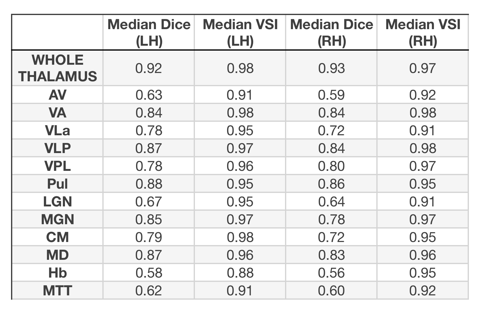
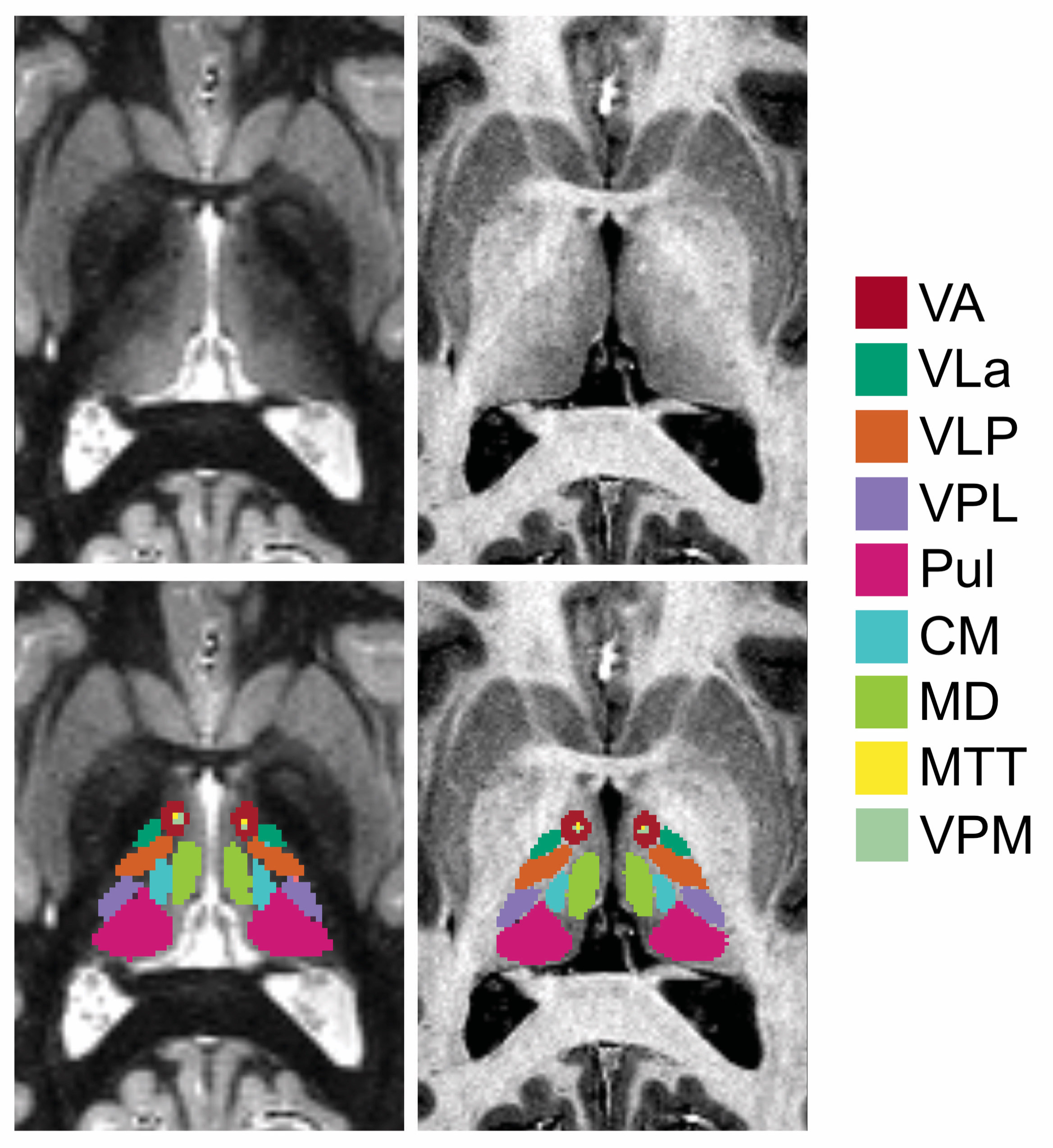
Segmentation time for a single subject was ~ 1 hr on a 6-core 3.5 GHz Intel Xeon. Figure 1 shows the median dice coefficients and the median volume similarity indices for the different nuclei identified in the ME-MP2RAGE and WMn-MPRAGE. The high dice coefficients and the high volume similarity indices indicate that THOMAS works as well with ME-MP2RAGE as with the WMn-MPRAGE images. Fig. 2 shows the segmentation results of THOMAS on WMnMPRAGE and ME-MP2RAGE.DISCUSSION/CONCLUSION
The THOMAS algorithm can successfully segment thalamic nuclei in MP2RAGE images with essentially equivalent quality, when evaluated against WMn-MPRAGE. The high accuracy in segmentation may in part be due to the fact that the T1 weighted image (UNI) reconstructed from the ME-MP2RAGE sequence has improved B1+ uniformity as the transmit and receive coil heterogeneity effects are uniform across the different TIs and hence are nullified.Reliable automatic segmentation of the whole thalamus and its nuclei is critical to understanding how the thalamic nuclei and their associated structural and functional connectivity are altered in aging and disease as well as the role of thalamic nuclei in human cognition. It may also play an important role for treatment planning, e.g. in thalamic ablation for essential tremor or deep-brain stimulation for a variety of disorders. Since MP2RAGE sequence is increasingly being used in 7T studies, the successful demonstration of thalamic nuclei delineation suggests wider applicability of the method to more studies.Acknowledgements
Research support from NMSS PP-1901-33149References
1. Thalamus Optimized Multi Atlas Segmentation (THOMAS): fast, fully automated segmentation of thalamic nuclei from structural MRI. Su JH, Thomas FT, Kasoff WS, Tourdias T, Choi EY, Rutt BK, Saranathan M. Neuroimage. 2019 Jul 1;194:272-282.
2. Visualization of intra-thalamic nuclei with optimized white-matter-nulled MPRAGE at 7T. Tourdias T1, Saranathan M, Levesque IR, Su J, Rutt BK. Neuroimage. 2014 Jan 1;84:534-45.
3. MP2RAGE, a self bias-field corrected sequence for improved segmentation and T1-mapping at high field. Marques JP, Kober T, Krueger G, van der Zwaag W, Van de Moortele PF, Gruetter R. Neuroimage. 2010 Jan 15;49(2):1271-81.
4. Simultaneous Quantitative MRI Mapping of T1, T2* and Magnetic Susceptibility with Multi-EchoMP2RAGE. Metere R, Kober T, Möller HE, Schäfer A. PLoS One. 2017 Jan 12;12(1):e0169265.
Figures