2000
Qualitative and Quantitative Evaluation of Uniform Combined Reconstruction (UNICORN) for Improving Intensity Uniformity in 7T-MRI of the Brain1Siemens Medical Solutions USA, Inc., Rochester, MN, United States, 2Department of Radiology, Mayo Clinic, Rochester, MN, United States, 3Siemens Medical Solutions USA, Inc., Boston, MA, United States, 4Department of Radiology, Brigham and Women's Hospital, Boston, MA, United States
Synopsis
Receive non-uniformity is one of the challenges that impacts 7T MRI and should be addressed for improved interpretation of brain images. Furthermore, segmentation algorithms that perform volumetric analysis of brain structures often require uniform signal intensity throughout the brain volume to function effectively. Uniform combined reconstruction (UNICORN) was recently reported as a method to improve receive uniformity in 7T musculoskeletal MRI; however, it has not yet been applied to brain imaging. The purpose of this work is to apply UNICORN to brain imaging and quantitatively evaluate its efficacy in improving intensity uniformity in 7T-MRI of the brain.
Introduction
7T-MRI of the brain benefits from high resolution, improved susceptibility contrast, enhanced signal-to-noise-ratio (SNR), better contrast-to-noise-ratio (CNR), increased T1 and other advantages provided by the ultra-high field (UHF) (1,2). Receive non-uniformity (3) is one of the challenges that impacts 7T MRI and should be addressed for improved interpretation of brain images. Furthermore, segmentation algorithms that perform volumetric analysis of brain structures often require uniform signal intensity throughout the brain volume to function effectively (4,5). Uniform combined reconstruction (UNICORN) was recently reported as a method to improve receive uniformity in 7T musculoskeletal MRI (3); however, UNICORN has not yet been applied to brain imaging. The purpose of this work is to apply UNICORN to brain imaging and qualitatively/quantitatively evaluate its efficacy in improving intensity uniformity in 7T-MRI of the brain.Methods
9 subjects underwent 7T brain examination on a MAGNETOM Terra (Siemens Healthcare, Erlangen, Germany) under the guidelines of an Institutional Review Board. A single-channel transmit, 32-channel phased-array receive head coil (Nova Medical Inc., MA, USA) was used for imaging the subjects. A 2D turbo-spin-echo (TSE) sequence was used to acquire fluid-attenuated inversion recovery (FLAIR) and T2-weighted MRI images. Imaging parameters for the FLAIR MRI included: TE of 52ms, TR of 11.33s, receive bandwidth (BW) of 455Hz/pixel, inversion time (TI) of 2.5s, flip angle of 120° and voxel dimensions of 1.5mmx1.5mmx2mm. Imaging parameters for the T2-weighted MRI included: TE of 51ms, TR of 8.25s, BW of 240Hz/pixel, flip angle of 120° and voxel dimensions of 2mmx2mmx2mm. Both standard and UNICORN images were obtained for each series of acquired data by processing the multi-channel receive data using prototype reconstruction software that implements the algorithm described in (3).Metric Used for Assessing Global Intensity Uniformity
The intensity uniformity of each 3D volume was assessed quantitatively as follows: The images from the multi-slice 2D TSE sequence were combined to form a 3D volume of brain MRI. The 3D volume was divided into eight equal volume octants $$$O_i$$$ (i ranging from 1 to 8). The average value ($$$\mu_i$$$) of the intensities within each octant $$$O_i$$$ was computed. Then, the standard-deviation ($$$\sigma_\mu$$$) and average ($$$\mu_\mu$$$) of the $$$\mu_i$$$ values were computed. Finally, intensity uniformity of the 7T brain MRI volume was estimated globally as inter-octant intensity homogeneity (IOIH):
$$IOIH = 1-\frac{\sigma_\mu}{\mu_\mu}$$
A paired two-sample T-test was performed to analyze the difference between IOIH computed from unnormalized and UNICORN normalized images.
Results
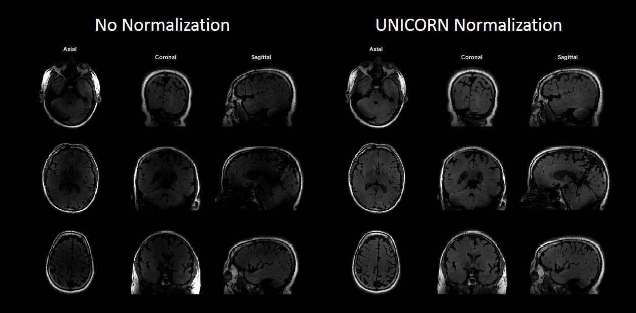
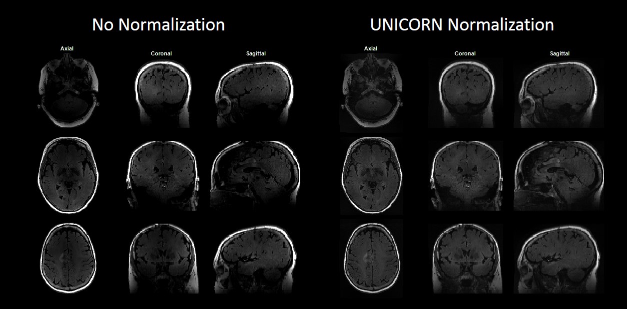
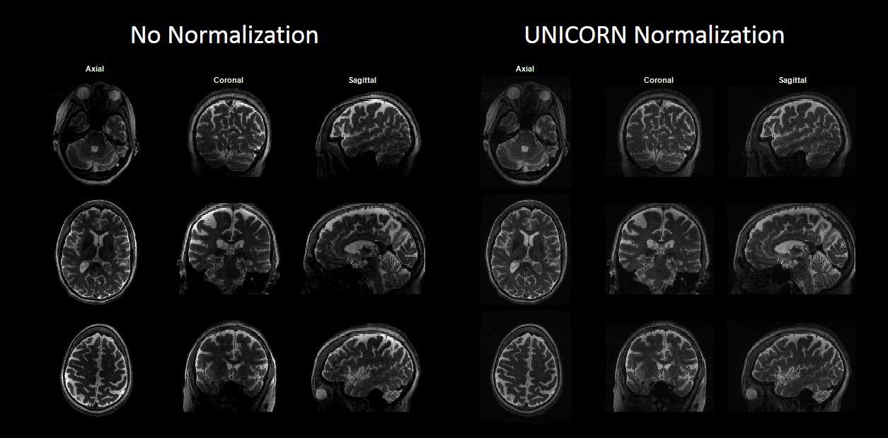
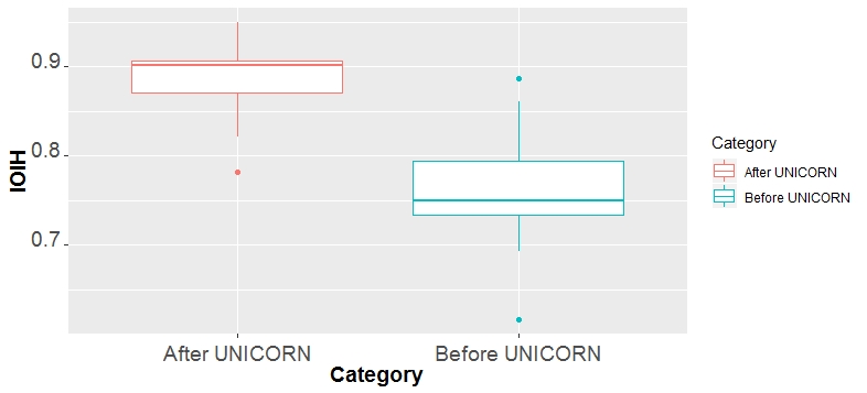
Figures 1 and 2 demonstrate the utility of UNICORN for improving uniformity of 7T brain FLAIR MRI in 2 representative subjects (data acquired in an axial orientation). Multiplanar reconstruction (MPR) of the images with no-normalization and with UNICORN normalization are shown to demonstrate through-slice profile and compare uniformity throughout the imaging volume. Figure 3 compares the uniformity between images with no-normalization and UNICORN normalization for a 7T brain T2-weighted MR data acquired in an axial orientation.Figure 4 shows the box plots of IOIH measured from the images with no-normalization and UNICORN normalization. The IOIH improvement achieved by UNICORN was statistically significant (p-value = 0.0001363).
Discussion
Receive-coil sensitivity profile depends on the shape, size and orientation of the coil and the sensitivity decreases quadratically with increase in distance from the coil. The decrease in receive-coil sensitivity with distance is more rapid at 7T or UHF compared to 3T or other lower main magnet field strengths creating higher intensity inhomogeneity (hypo-intensities at the center of field-of-view and hyper-intensities proximal to the coils) in a 7T coil-combined MR image. The absence of a reference “body coil” in the 7T MR system precludes the possibility of using a body-coil calibration-based intensity correction such as prescan normalize (PSN) often used at lower field strengths to address receive non-uniformity. In this work a data-driven coil-sensitivity estimation approach, UNICORN, was used to improve the intensity uniformity in 7T-MRI of the brain. The efficacy of UNICORN in improving uniformity was visually assessed and it was observed that UNICORN addressed the issue of hypo-intensity in the central regions of the field-of-view to a large extent.The improvement in uniformity achieved by UNICORN was also evaluated quantitatively using the IOIH metric. The metric measures variation in intensity across the eight octants in a 3D volume and provides a high uniformity score (close to 1) for the volumes with less inter-octant intensity differences and a lower uniformity score for the volumes with higher inter-octant intensity differences. The IOIH metric provides a global measure of uniformity. One limitation of a global uniformity measure (estimated using IOIH or otherwise) is that global metrics do not measure uniformity for each brain tissue category separately. Analyzing uniformity in each individual tissue class separately requires segmentation, which was beyond the scope of this work.
Conclusion
The UNICORN algorithm improved uniformity of 7T-MRI of the brain both visually (or qualitatively) and quantitatively as estimated by the inter-octant intensity homogeneity (IOIH) metric.Acknowledgements
No acknowledgement found.References
1. Karamat MI, Darvish-Molla S, Santos-Diaz A. Opportunities and challenges of 7 tesla magnetic resonance imaging: A review. Crit. Rev. Biomed. Eng. 2016. doi: 10.1615/CritRevBiomedEng.2016016365.
2. Moser E, Stahlberg F, Ladd ME, Trattnig S. 7-T MR-from research to clinical applications? NMR Biomed. 2012. doi: 10.1002/nbm.1794.
3. Chebrolu VV, Kollasch PD, Deshpande V, et al. Uniform combined reconstruction of multichannel 7T knee MRI receive coil data without the use of a reference scan. J. Magn. Reson. Imaging 2019. doi: 10.1002/jmri.26691.
4. Schindler S, Schreiber J, Bazin PL, Trampel R, Anwander A, Geyer S, Schönknecht P. Intensity standardisation of 7T MR images for intensity-based segmentation of the human hypothalamus. PLoS One 2017. doi: 10.1371/journal.pone.0173344.
5. Uwano I, Kudo K, Yamashita F, Goodwin J, Higuchi S, Ito K, Harada T, Ogawa A, Sasaki M. Intensity inhomogeneity correction for magnetic resonance imaging of human brain at 7T. Med. Phys. 2014;41:022302. doi: 10.1118/1.4860954.
Figures