1995
Altered network efficiency is associated with mood disturbance in adults with major depressive disorder1Department of Radiology, Second Affiliated Hospital of Zhejiang University School of Medicine, Hangzhou, China, 2Department of Psychiatry, Tongde Hospital of Zhejiang Province, Hangzhou, China
Synopsis
To better understand the underlying mechanisms for emotional disturbances in patients with major depressive disorder (MDD), we used structural brain connectivity analysis to investigate the differences in global and local network organization of MDD and healthy controls. Our results demonstrated that both global and local efficiency of patients with MDD were significantly decreased due to depressive symptoms, and that higher depression severity, anxiety somatization and cognitive disturbance were significantly associated with decreased network efficiency. These results indicated that brain network analysis is a useful tool to link psychological disorders with their underlying anatomical substrate.
Purpose
With a lifetime prevalence of more than 15%, major depressive disorder (MDD) causes considerable impairment and substantially worsens mean health scores when comorbid with other chronic diseases. While early neuroimaging studies focused on localizing the depression related symptoms to specific brain regions, recent frameworks emphasize that disrupted brain network might be the pathogenesis of mood disturbance in MDD. Neural systems that are supportive for emotion processing and mood regulation are important to MDD. Considering the identification of those systems might be helpful for guiding treatment choice, we therefore aim to investigate the structural network alterations in patients with MDD when compared to healthy subjects, and their relations with depression severity.Materials and methods
Participants 26 MDD patients (39 ± 13 years old) experiencing moderate or severe depressive episode according to the DSM-IV-TR criteria1 were recruited. 13 age and sex matched healthy controls (HCs) (33 ± 7 years old) without any neurological or neuropsychological disorders were recruited.Image acquisition Diffusion weighted images along 30 gradient directions with a b-value of 1000 were acquired using a single-shot EPI sequence (Siemens Trio 3-tesla system; Erlangen, Germany). High-resolution three-dimensional structural images were acquired by using a T1-weighted gradient echo sequence at a voxel size of 1 mm3 isotropic, parallel to the anterior commissure–posterior commissure (AC–PC) line and covering the whole brain.
Post-processing T1-weighted images were segmented into 90 brain regions according to AAL atlas. Whole-brain tractography was obtained using Diffusion Toolkit (trackvis.org/dtk/). Structural network was estimated by counting the number of fiber tracts traversed any pairs of two regions.
Network analysis Global network topological properties, including global efficiency and local efficiency, and regional network measures were obtained using the Brain Connectivity Toolbox2.
Statistical analysis Two sample permutation test was used to test the difference in network measures between patients with MDD and HC. The associations between network measures and the depression level of patients, measured by Hamilton Depression Rating Scale (HAMD), were investigated using Spearman’s rank correlation. All the p-values were corrected for multiple comparisons using FDR.
Results
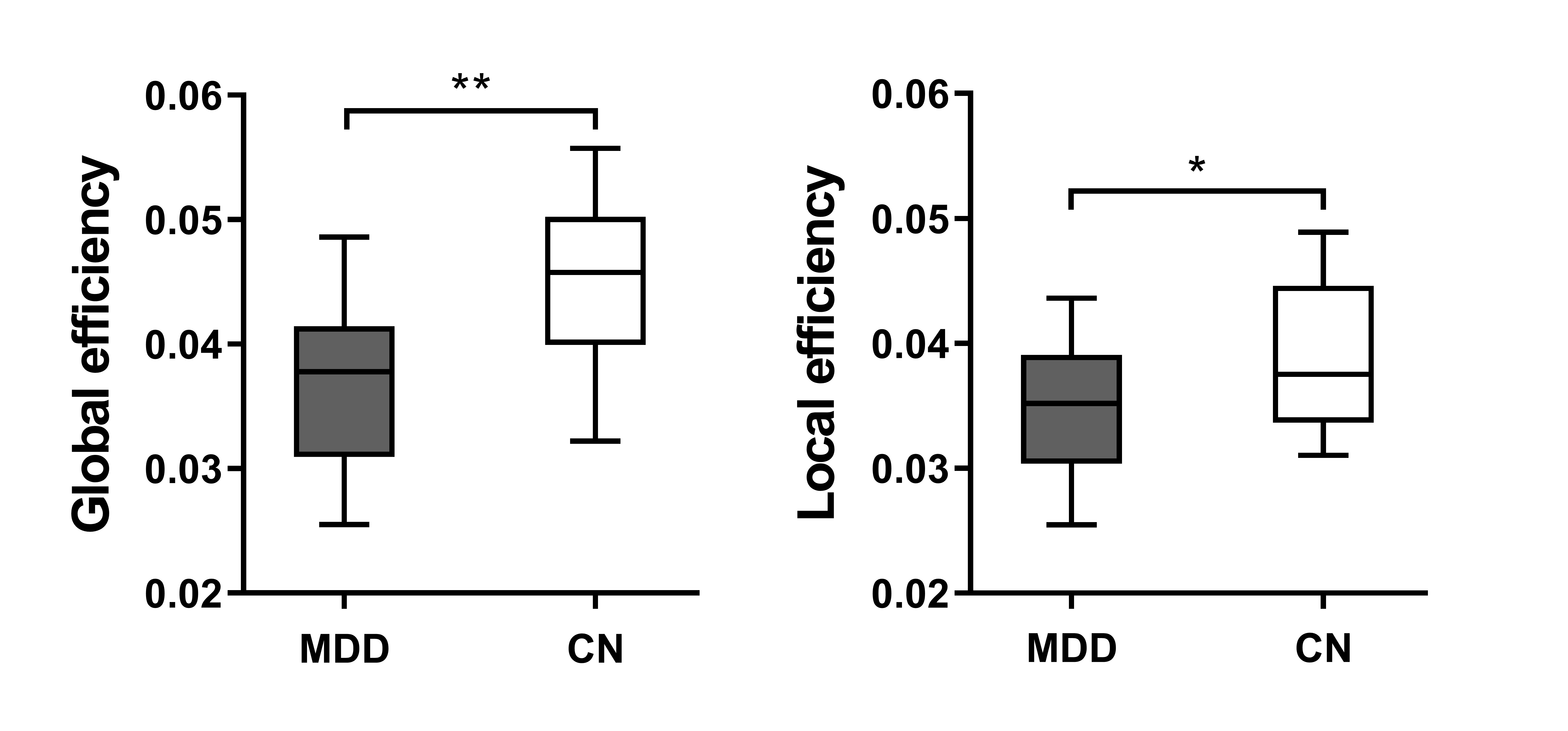
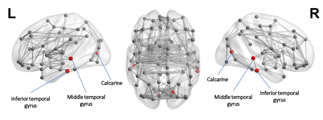
Both patients with MDD and HCs had significantly larger clustering coefficient and relatively same characteristic path length as compared to random network, which means they both demonstrated a typical small-world network. When compared to HCs, significantly higher characteristic path length (p = 0.002) was observed in patients with MDD. Furthermore, MDD patients also showed significantly decreased global (p < 0.001) and local efficiency (p = 0.039), as shown in Figure 1. We also investigated the differences in regional network features between two cohorts. Significantly lower nodal efficiency was found in calcarine fissure (p = 0.003), middle temporal gyrus (p = 0.003) and inferior temporal gyrus (p = 0.034) (See Figure 2). Most importantly, we found that the characteristic path length of MDD patients was associated with weight loss (p = 0.049) in MDD patients. Also, the global efficiency, which is an indicator of cognitive function, was negatively correlated with total HAMD score (p = 0.041), anxiety somatization (p = 0.002) and cognitive disturbance (p = 0.045) in patients with MDD.Discussion
The linkage between network changes and pathological states of brain has been established in many neuropsychiatric diseases, such as Alzheimer’s disease3, schizophrenia4 and epilepsy5. In the current study, the patient with MDD had significantly lower global and local efficiency, indicating that the long- and short-range connections among regions were disrupted when compared to HCs, potentially related to the emotional disorders of these patients 6. As past evidence suggested that cortical disconnection syndrome might be the underlying neurophysiological mechanism in MDD, Our results showed certain consistency with previous studies7 which have shown abnormal prefrontal cortico-subcortical white matter connectivity between regions supporting emotion regulation in MDD patients. In addition to global network changes, we successfully identified regions that are particularly vulnerable to the disease insult, namely calcarine fissure, middle temporal gyrus and inferior temporal gyrus. This finding is in line with past studies which demonstrated that patients with depressive symptoms exhibited greater atrophy in temporal lobe8. More importantly, we found that higher global efficiency was associated with lower depression level, as measured by HAMD, anxiety somatization and cognitive disturbance. Considering that HAMD indicates depression severity and is considered a guide to evaluate recovery9, the association between network efficiency and HAMD that we observe may serve as an evidence for the notion that disrupted network connection and decreased mood regulation ability might be the causes to emotional disturbance in MDD.Conclusion
Our findings showed that patients with MDD exhibit disrupted global and local network topological organization when compared to HCs. More importantly, this altered network configuration with impaired regional information exchange ability may contribute to the depressive symptoms in MDD.Acknowledgements
No acknowledgement found.References
1. American Psychiatric Association. Diagnostic and Statistical Manual of Mental Disorders. American Psychiatric Association; 2013.
2. Bullmore E, Sporns O. Complex brain networks: graph theoretical analysis of structural and functional systems. Nat Rev Neurosci 2009;10:186–98.
3. Nir T, Jahanshad N, Jack CR, et al. Small world network measures predict white matter degeneration in patients with early-stage mild cognitive impairment. In: 2012 9th IEEE International Symposium on Biomedical Imaging (ISBI). IEEE; 2012:1405–8.
4. Liu Y, Liang M, Zhou Y, et al. Disrupted small-world networks in schizophrenia. Brain 2008;131:945–61.
5. Reijneveld JC, Ponten SC, Berendse HW, et al. The application of graph theoretical analysis to complex networks in the brain. 2007;118:2317–31.
6. Gaete JM, Bogousslavsky J. Post-stroke depression. Expert Rev Neurother 2008;8:75–92.
7. Li L, Ma N, Li Z, et al. Prefrontal white matter abnormalities in young adult with major depressive disorder: a diffusion tensor imaging study. Brain Res 2007;1168:124–8.
8. Shimada H, Park H, Makizako H, et al. Depressive symptoms and cognitive performance in older adults. J Psychiatr Res 2014;57:149–56.
9. Williams JB. Standardizing the Hamilton Depression Rating Scale: past, present, and future. Eur Arch Psychiatry Clin Neurosci 2001;251 Suppl 2:II6-12.
Figures