1992
The OCD symptoms affect to abnormal functional activity and connectivity at caudate nucleus during memory retrieval, and lead to GM volume changes
Shin-Eui Park1, Gwang-Woo Jeong2, and Chulhyun Lee1
1Center for research equipment, Korea Basic Science Institue, Cheongju, Republic of Korea, 2Department of Radiology, Medical school, Chonnam National University, Gwangju, Republic of Korea
1Center for research equipment, Korea Basic Science Institue, Cheongju, Republic of Korea, 2Department of Radiology, Medical school, Chonnam National University, Gwangju, Republic of Korea
Synopsis
For verifying how the OCD symptom affects to abnormal functional activity and connectivity at Cd during memory processing, and whether leading to brain volume changes in direct or indirect, we performed multi study combined with fMRI and VBM study. Twenty OCD patients and 20 controls took part in VBM and fMRI studies with explicit memory task. The functional and morphological profiles of Cd could be emphasized to abnormal processing of explicit memory by OCD symptoms. It puts forward a possibility that focal functional connectivity and brain volume changes may be affected by lasting OCD symptoms, and leading to memory dysfunction.
Introduction
The caudate nucleus (Cd) is one of the structures that make up the corpus striatum, which is a component of the basal ganglia. This brain area is one of the brain structures which involved in the memory system and functions as part of the cortico–basal ganglia–thalamic loop. The patients with obsessive compulsive disorder (OCD) have shown cognitive defect and neurofunctional abnormality through various clinical and functional studies. It can be assumed that Cd may be dysfunctional in persons with OCD, in that it may perhaps be unable to properly regulate the transmission of information during memory processing between the thalamus and the prefrontal cortex. Therefore, for verifying how the OCD symptom affects to abnormal functional activity and connectivity at Cd during memory processing, and whether leading to brain volume changes in direct or indirect, we performed multi study combined with fMRI and VBM study.Methods
Forty right-handed subjects included twenty patients with OCD (mean age, 29.4±9.9 years) who are diagnosed by a psychiatrist using the DSM-IV-TR and 20 healthy controls (mean age, 29.7±8.3 years) with no history of neurological or psychiatric illness. The duration of patient’s illness was 5.8±4.7 years. All the participants underwent the clinical interviewed by using Yale-Brown Obsessive-Compulsive Scale, Hamilton Depression Scale 17 and Hamilton Anxiety Scale. The difference of symptom severity in both groups was analyzed by Mann-Whitney U test. The activation paradigm for memory processing consisted of the following cycle: rest (14s), first encoding (18s), rest (14s), first retrieval (18s), rest (14s), second encoding (18s), rest (14s), second retrieval (18s), and rest (14s). MR scanning was performed on a 3 Tesla Magnetom Verio MR Scanner (Siemens Medical Solutions, Erlangen, Germany) with an 8-channel head coil. Anatomical T1-weighted images were acquired using a three-dimensional magnetization-prepared rapid acquisition gradient-echo sequence with repetition time (TR)/echo time (TE)= 1900/2.35 ms, field-of-view (FOV)= 22×22 cm2, matrix size= 256×256, number of excitation (NEX)= 1 and slice thickness= 1 mm. functional images were acquired from a total of 25 axial slices parallel to an anterior commissure to posterior commissure line using a gradient-echo echo planner image (GRE-EPI) with the follow parameters: TR/TE= 2000/30 ms, flip angle= 90°, FOV= 22×22 cm2, metrix size= 64×64, NEX= 1, and slice thickness= 5mm without a slice gap. The 3D T1 image data were processed by SPM8 with DARTEL tool. The fMRI data were processed by using SPM12 and CONN software.Results
In fMRI results, patients with OCD showed higher activities in the right/left inferior temporal gyrus (ITG), medial prefrontal cortex (MPFC), dorsolateral prefrontal cortex (DLPFC) and anterior cingulate cortex (ACC) compare to healthy controls in memory encoding (uncorrected P<0.001) (figure 1a). The Cd, DLPFC and ITG had a positively connected each other in OCD patients. However, the Cd connected with ACC and right ITG connected with left ITG positively in healthy controls (uncorrected P<0.001) (figure 2a). In memory retrieval, Cd is observed as higher activity in OCD patients (uncorrected P<0.001) (figure 1b). This Cd has composed positive connectivity with DLPFC and vmPFC in OCD patients, but negative connectivity in healthy controls (uncorrected P<0.001) (figure 2b). In VBM, OCD patients showed a significant increase in gray matter (GM) volumes: cerebellum, DLPFC, orbitofrontal cortex (OFC), Hippocampus, Cd and TIG and white matter volumes: DLPFC (FWE P<0.05) (figure 3). The GM volume in Cd and OFC of patients with OCD showed positive correlation with HAMA and Y-BOCS (r=0.458, p=0.042; r=0.525, p=0.016) (figure 4). The BOLD signal changes in Cd of patients with OCD showed positive correlation with Y-BOCS (r=0.478, p=0.033) (figure 5). In summary, the lasting psychiatric symptoms with OCD more than five years may be closely associated with neurofunctional and morphological changes, give rise to abnormal explicit memory processing.Conclusion
This finding will be helpful to understand the neuroanatomical and functional mechanism for explicit memory processing in relation to OCD symptoms. In particular, it puts forward a possibility that focal functional connectivity and brain volume changes may be affected by lasting OCD symptoms, and leading to memory dysfunction. The functional and morphological profiles of Cd could be emphasized to abnormal processing of explicit memory by OCD symptoms. Furthermore, it will be enhancing the diagnostic accuracy for OCD as additional information on the brain functional and cerebral volume charges.Acknowledgements
References
1. Yager LM, et al, Neuroscience. 2015;301:529-41. 2. Jang IS, et al. Neuroreport. 2017;28:1-9.Figures
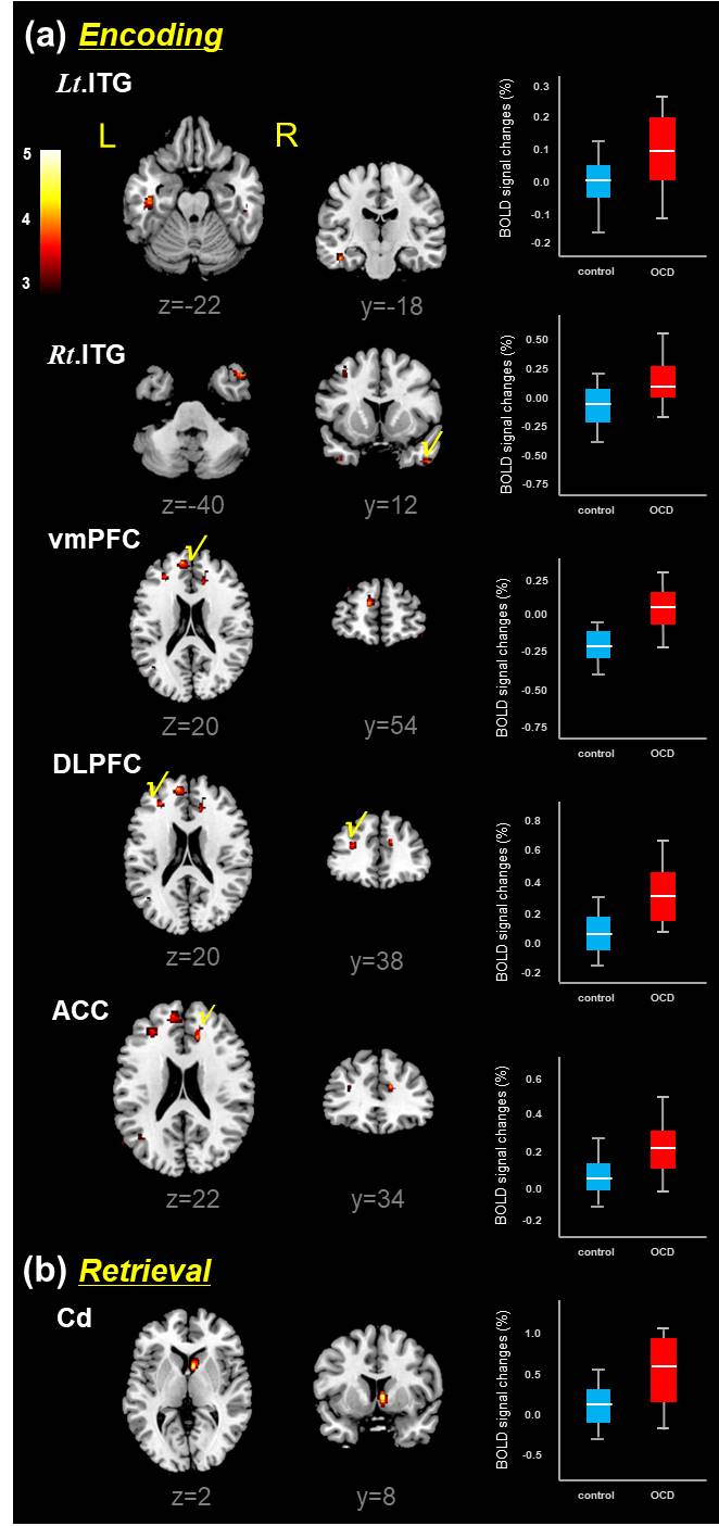
Brain
areas predominantly activated in
patients with OCD relative to healthy controls during memory task
(encoding
epoch (a) and retrieval epoch (b)), which resulted from the
two-sample t-test (uncorr., P<0.001). Box-plots show
the medians, interquartile ranges, highest and lowest values for BOLD signal
intensities of corresponding brain areas. The color bar represents the t-value.
ITG, inferior temporal gyrus; vmPFC, ventromedial prefrontal cortex;
DLPFC, dorsolateral prefrontal cortex; ACC, anterior cingulate cortex; Cd, caudate nucleus. L= left; R= right.
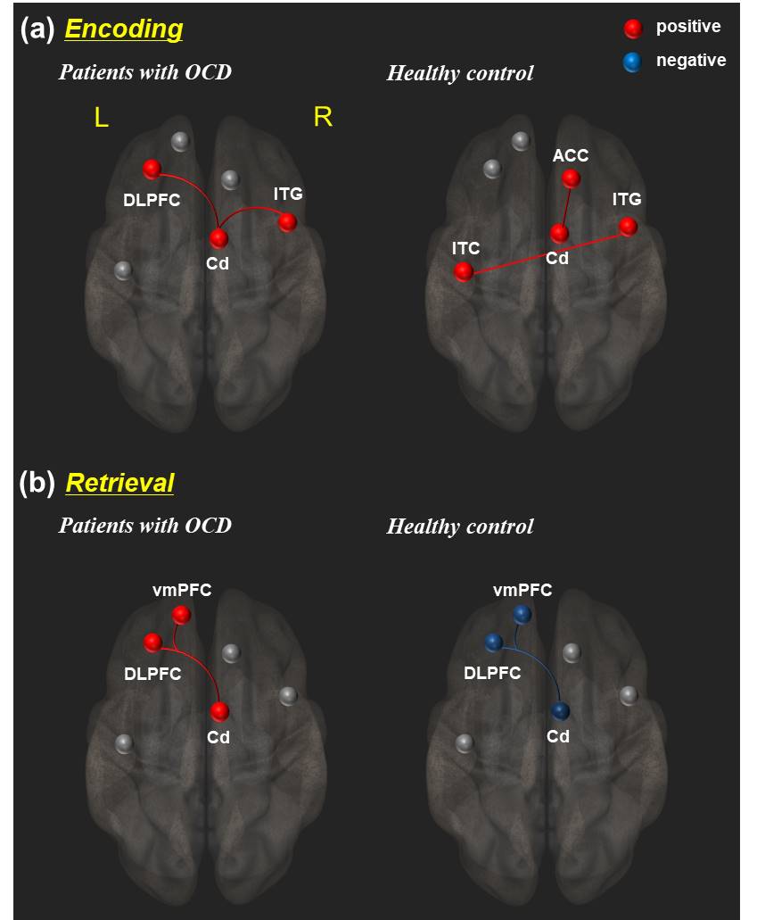
Different
brain connectivity
patterns in
the
OCD
patients and healthy controls during memory task (encoding epoch (a) and
retrieval epoch (b)). Red
lines represent increased
functional
connectivity strength
and blue
lines represent decreased
functional
connectivity strength
(uncorrected p<0.05). Details
can be found at Table
3. DLPFC, dorsolateral prefrontal cortex; ITG, inferior temporal gyrus; Cd,
caudate nucleus;
ACC, anterior cingulate cortex; vmPFC,
ventromedial prefrontal cortex. L=left; R=right.
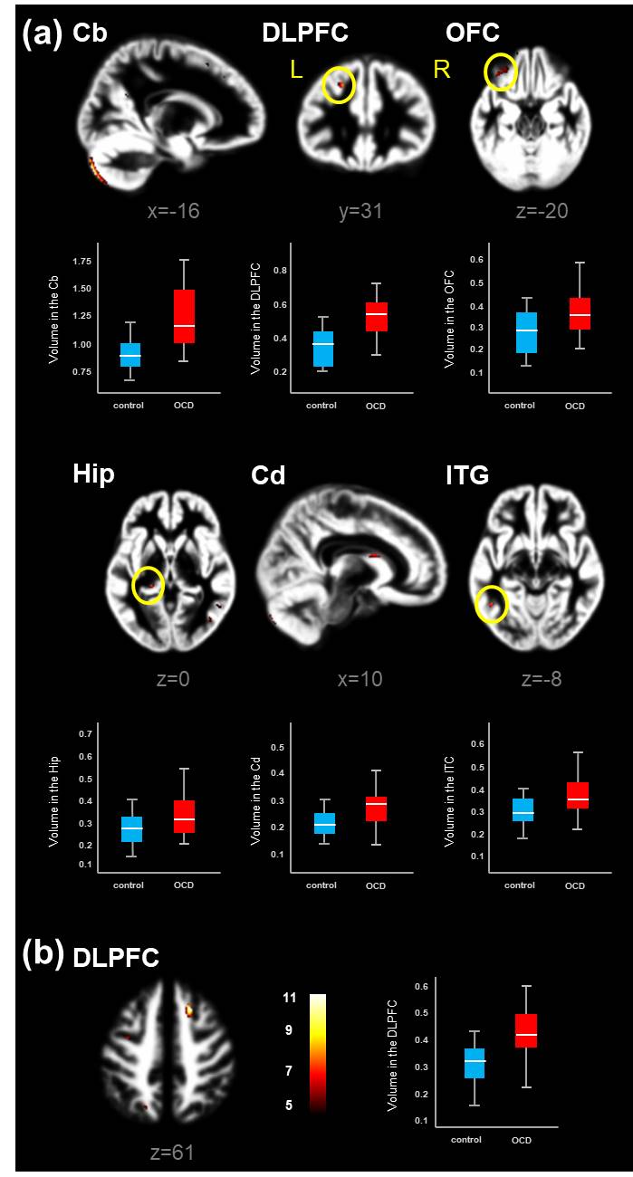
Brain
areas showing significant increased gray matter (a) and white matter (b)
volumes in patients with OCD relative to healthy controls, which resulted from
the two-sample t-test (FEW, P<0.05). Box-plots show the medians,
interquartile ranges, highest and lowest values for regional volumes (mL) of
corresponding brain areas. Cb,
cerebellum; DLPFC, dorsolateral prefrontal cortex; OFC, orbitofrontal cortex;
Hip, hippocampus, Cd, caudate nuclus;
ITG, inferior temporal gyrus. L= left; R= right.
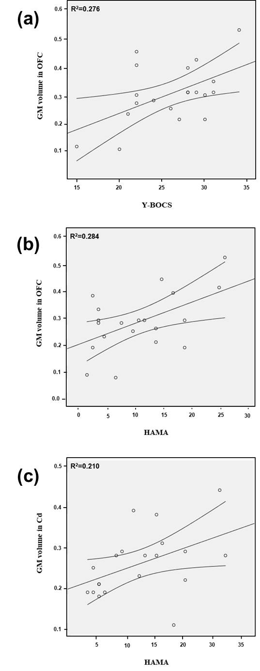
The
correlation of the symptom severity and gray matter (GM) volume changes in
patients with OCD using correlation analysis: Y-BOCS scores vs. GM volumes in
orbitofrontal cortex (OFC) (a), and HAMA scores
vs.
GM volumes in OFC (b) and caudate
nucleus (Cd) (c)
The
curved line bands mean 95%
confidence intervals.
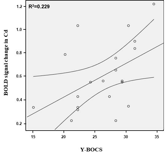
The
correlation between the Y-BOCS scores and BOLD signal changes in caudate nuclus
(Cd) of patients with OCD using correlation analysis
(r=0.478, p=0.033).
The curved line bands mean 95% confidence intervals.