1989
Temporal variability of local brain activity in medication-free patients with Obsessive Compulsive Disorder1Huaxi MR Research Center (HMRRC), Functional and molecular imaging Key Laboratory of Sichuan Province, Department of Radiology, West China Hospital, Sichuan University, Chengdu 610041, China, Chengdu, China
Synopsis
In current study, we use static and dynamic amplitude of low-frequency fluctuation / fractional low-frequency fluctuation (ALFF/fALFF) to assess the local brain activity on patients with obsessive-compulsive disorder (OCD). We found that both static and dynamic ALFF/fALFF can detect certain cerebral region with abnormal activity, however, dynamic ALFF/fALFF can demonstrate additional brain regions that are ignored by static ALFF/fALFF. In addition, we found that there is significant correlation between cerebellum activity and clinical scale, which suggested the crucial role of cerebellum in the pathophysiology of OCD.
Background
Amplitude of low-frequency fluctuation / fractional low-frequency fluctuation(ALFF/fALFF) which is a commonly used parameter extracted from resting-state fMRI(rs-fMRI) can reflect the intensity of spontaneous neural activity. However, as the brain is a highly dynamic system 1 and traditional ALFF/fALFF can not capture the complex and dynamic changes of brain activity, recently, a new parameter called dynamic ALFF/fALFF(d-ALFF/d-fALFF) was used to reflect brain activity changes in terms of temporal variability in several psychiatric disorders 2,3.Obsessive-compulsive disorder (OCD) is a common mental disorder affects approximately 2%–3% of the population and characterized by invasive thoughts and repetitive behavior 4. However, few studies have explored the cerebral dynamic changes in OCD, so in current study, we aimed to demonstrate whether OCD patients exist temporal variability alternation of local brain activity using d-ALFF/d-fALFF and we also combine the traditional static ALFF/fALFF(s-ALFF/s-fALFF) to help fully understand the characteristic change of cerebral intrinsic activity in OCD.Methods
A total of 53 medication-free OCD patients (age 30.26 ± 8.08) and 52 sex and age matched HCs (age 28.48 ± 11.29) were recruited in current study (Table 1). We used Yale-Brown Obsessive Compulsive Scale (Y-BOCS) to assess severity of OCD symptoms and used Hamilton Anxiety Scale (HAMA) and Hamilton Depression Scale (HAMD) to assess depression level and anxiety.All the subjects were scanned by a 3-Tesla GE MRI system.The T1 and rs-fMRI images were obtained for each subject and preprocessed using DPARSF software(http://www.restfmri.net). The static and dynamic ALFF/fALFF maps were estimated by DPABI toolbox (http://www.rfmri.org/dpabi). For s-ALFF/s-fALFF, we calculated the mean value during the whole session for each voxel. For d-ALFF/d-fALFF, we applied sliding window technique with a window size of 50 TRs (100 seconds) basing on the “rule of thumb,” which is 1/fmin of data should be equal or less than the length of window 5.The standard deviation (SD) maps of ALFF/fALFF for each subject across 141 windows were calculated which were then used to assess the temporal variability of local brain activities. Then static maps and SD maps were standardized and smoothed.
We used two-sample t-tests with FWE correction (p corr < 0.05 at cluster level, p uncorr <0.001 at peak level) to demonstrate brain regions with significant differences between the two groups. In addition, we calculated the associate between clinical symptom severity and the regions with abnormality of cerebral activity using Pearson correlation.
Results
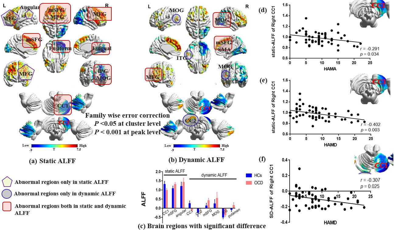
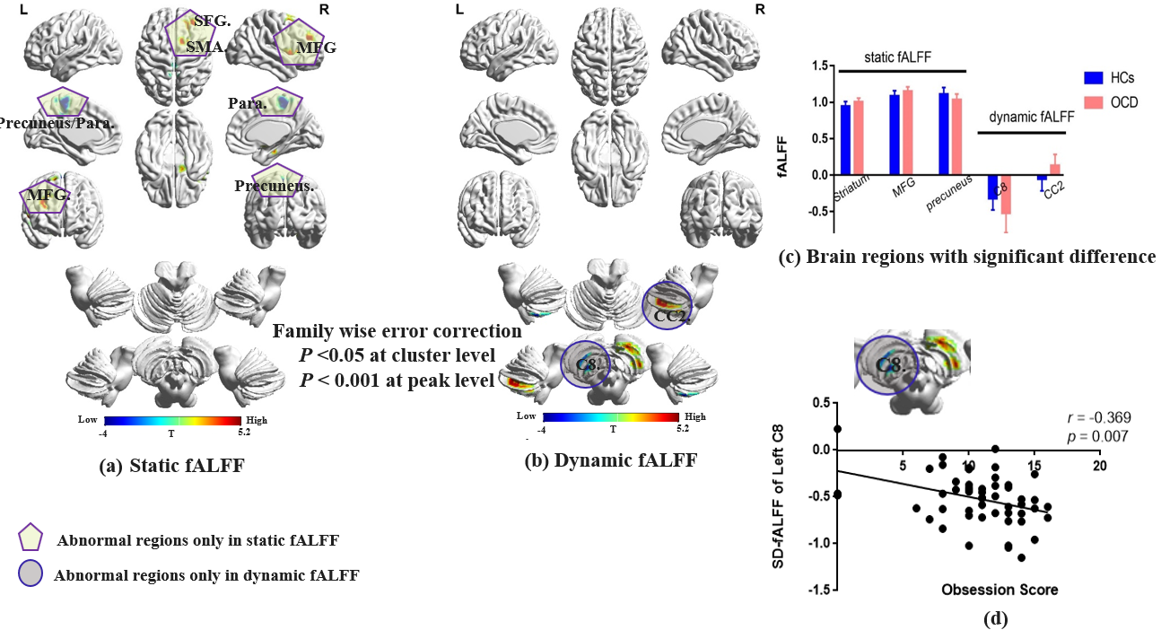
1. Static and dynamic ALFF: Compare to HCs, OCD patients showed both higher static and dynamic ALFF in bilateral medial superior frontal gyrus (mSFG), lower static and dynamic ALFF in right middle occipital gyrus (MOG), right crus I of cerebellum (CC1)and fusiform. The abnormalities of increased intrinsic activity in left angular in OCD patients was only detected by s-ALFF. And the abnormalities of increased temporal variability in left MOG, left inferior temporal gyrus (ITG) and crus II of cerebellum (CC2) were only detected by d-ALFF (figure 1 and table 2).2. Static and dynamic fALFF: Group comparison of s-fALFF mainly located abnormalities in the cerebrum including increased intrinsic activity in right middle frontal gyrus (MFG), superior frontal gyrus (SFG)and decreased intrinsic activity in bilateral precuneus/paracentral lobule in OCD patients. Group comparison of d-fALFF mainly located abnormalities of increased temporal variablity in right CC2 and decreased temporal variablity in left cerebellum lobule VIII (C8) (figure 2, and table 2).
3. Both the s-ALFF and d-ALFF of right CC1 correlated negatively with HAMD, and s-ALFF of right CC1 correlated negatively with HAMA. Besides, d-fALFF of left C8 correlated negatively with obsession score (figure 1-2).
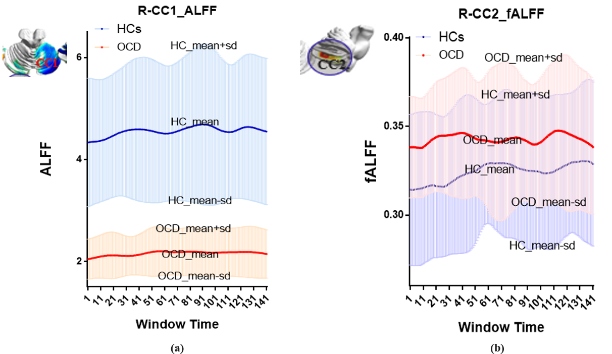
4. In order to show the specific changes of ALFF/fALFF across all Windows for each group, we plot out the mean and mean_SD curve of two regions in right CC1 which showed decreased temporal variability in OCD patients, and right CC2 which showed increased temporal variability in OCD patients as shown in figure 3.
Discussion & Conclusion
In this study, we found that OCD patients bears cerebral regional abnormalities in left MOG /ITG/CC2 which were only detected by d-ALFF and abnormalities in right CC2 and left C8 were only detected by d-fALFF. Thus, we proposed that d-ALFF/d-fALFF can detect additional cerebral activity difference between the two groups that may ignored by traditional s- ALFF/s-fALFF.The abnormalities of right CC1/MOG and bilateral mSFG/MFG were detected by both static and dynamic ALFF, indicating these regions have both regional activity amplitude abnormalities and temporal variability abnormalities in OCD patients.
In addition, we found abnormality in cerebellum in OCD patients with both static and dynamic parameters, and more importantly, the significant association between clinical scales and activity in certain regions of cerebellum indicate the critical role of this structure in the pathophysiology of OCD which deserves further investigation.
Taken together, our results suggest the combination of static and dynamic ALFF/fALFF can help to detect regional brain intrinsic activity in a more comprehensive way by considering both the amplitude and temporal variability of the BOLD signal. And this may be particular useful in mental disorders whose neural mechanism is still largely unknown.
Acknowledgements
This study was supported by National Nature Science Foundation (Grant NO. 81671669), Science and Technology Project of Sichuan Province (Grant NO. 2017JQ0001).References
1. Hutchison RM, Womelsdorf T, Gati JS, et al. Resting-state networks show dynamic functional connectivity in awake humans and anesthetized macaques. Human brain mapping. 2013; 34(9):2154-2177.
2. Fu Z, Tu Y, Di X, et al. Characterizing dynamic amplitude of low-frequency fluctuation and its relationship with dynamic functional connectivity: An application to schizophrenia. Neuroimage. 2018; 180:619-631.
3. Jiao L , Xujun D , Qian C , et al. More than just statics: temporal dynamics of intrinsic brain activity predicts the suicidal ideation in depressed patients[J]. Psychological Medicine. 2018;1-9.
4. Milad M R, Rauch S L. Obsessive-compulsive disorder: beyond segregated cortico-striatal pathways[J]. Trends in Cognitive Sciences. 2012; 16(1):0-51.
5. Leonardi N, Ville D V D. Erratum to “On spurious and real fluctuations of dynamic functional connectivity during rest’’[J]. Neuroimage. 2014; 104.
Figures
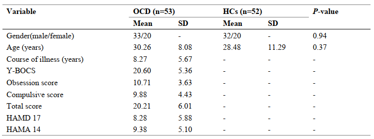
Table 1. Demographic and clinical characteristics of the participants. OCD, Obsessive-compulsive disorder; HCs, Healthy Control subjects; SD, standard deviation; Y-BOCS, Yale-Brown Obsessive-Compulsive Scale; HAMA, Hamilton Anxiety Scale; HAMD, Hamilton Depression Scale.
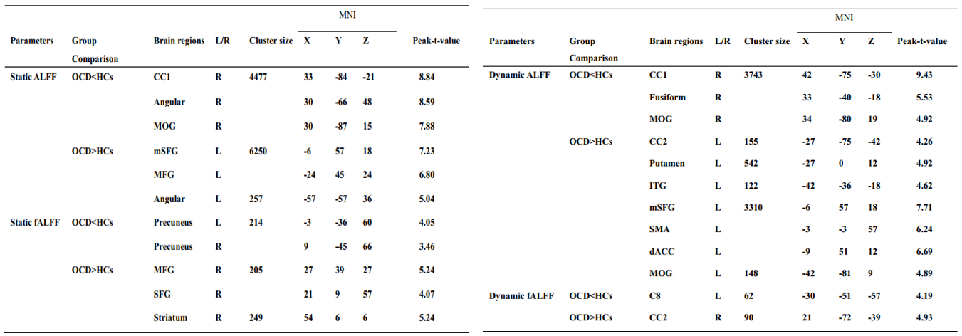
Table 2. Brain regions with significant difference between OCD and HCs. CC1, cerebellum crus I; MOG, middle occipital gyrus; mSFG, medial superior frontal gyrus; MFG, middle frontal gyrus; SFG, superior frontal gyrus; CC2,cerebellum crus II; ITG, inferior temporal gyrus; SMA, supplementary motor area; dACC, dorsal anterior cingulate cortex; C8, cerebellum lobule VIII.