1981
Evaluating the Role of Bed Nucleus of Stria Terminalis in Anorexia Nervosa Restricting Type1Radiology, Penn State College of Medicine, Hershey, PA, United States, 2Molecular Biology, Penn State College of Medicine, Hershey, PA, United States, 3Psychiatry, Penn State College of Medicine, Hershey, PA, United States
Synopsis
There is growing evidence for the contribution of neurocircuitry regulating emotion, as well as appetite and body weight, in the etiology of anorexia nervosa (AN). The high prevalence of anxiety disorders in AN implicates a critical node in the neurocircuitry of anxiety. The bed nucleus of the stria terminalis (BNST) modulates not only responses to anxiogenic (versus fear) situations, but mediates anxious temperament and feeding as well, through efferent connections to the lateral hypothalamus. In this study, we sought to identify if there is significant functional and structural changes of BNST in AN related to food.
Introduction
Although the neuropathological mechanisms underlying anorexia nervosa (AN) are still unknown, there is growing evidence for the contribution of neurocircuitry regulating emotion, as well as appetite and body weight, in the etiology of anorexia. The high prevalence of trait anxiety and anxiety disorders in AN implicates a critical node in the neurocircuitry of anxiety [1-4]. Anorexia is found predominately among females, and anxiety disorders are more prevalent in females. The sexually dimorphic bed nucleus of the stria terminalis (BNST) modulates not only responses to anxiogenic (versus fear) situations, but also mediates anxious temperament [5] and feeding, through efferent connections to the lateral hypothalamus [6]. BNST volume decrease due to cell loss occurs around or after puberty [7]. However, no studies on BNST structure or functions have been reported in AN. In this study, we sought to identify if there is significant functional and structural changes of BNST in AN related to food.Methods
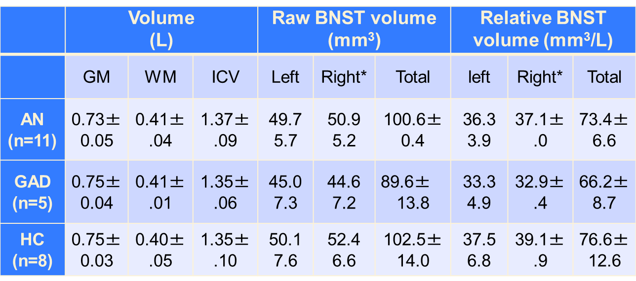
12 AN, 5 general anxiety disorder (GAD), and 8 healthy control (HC) subjects participated in the study (Table 1). All subjects were right-handed female teenagers. All subjects and/or guardians gave written informed consent, which was approved by the local institutional review board. BMI, STAI anxiety level, and taste functions (intensity and pleasantness of sweet and salty tastes in a scale of 0-3) were acquired before MRI. fMRI paradigms: During the gustatory stimulation fMRI paradigms, either 0.12 ml of a milkshake or 0.045 M NaCl was pseudorandomly delivered to the anterior portion of the subject’s tongue for sweet (high-calorie) and salty tastes. Subjects were trained to follow visual cues, indicating when to expect a tastant, hold it in the mouth, and swallow. During visual stimulation fMRI, the subjects expected and viewed either negative or neutral pictures. After scanning, subjective ratings of overall/peak anxiety levels during each fMRI paradigm in a scale of 0-100 were collected. fMRI was conducted on a Siemens Prisma Fit 3T scanner with a 64-channel head/neck coil and BOLD-sensitive T2*-weighted EPI in 3×3×4 mm3 resolution. In addition, whole brain was scanned with T1-weighted MPRAGE method in 1 mm3 resolution. fMRI data processing and analysis: fMRI data was processed with general linear model in SPM12. From the two taste paradigms, activation maps responding to expecting and holding the tastants in mouth were generated for each taste modality (milkshake and NaCl). From the visual paradigm, activation maps responding to expecting and viewing photos were generated for each image modality (negative and neutral). Group level activation maps were generated with one sample t-tests and cross-group comparisons between age-matched subjects were conducted with ANCOVA with age as a covariate. Morphological data processing and analysis: One AN subject’s morphologic image was missing. Voxel-based morphometry analysis was conducted with SPM12. The ROI analysis was based on manual segmentation of BNST using MRIcron. The absolute volume of BNST and its relative volume after correction by the intra-cranial volume (ICV) were used for cross-group comparisons.Results
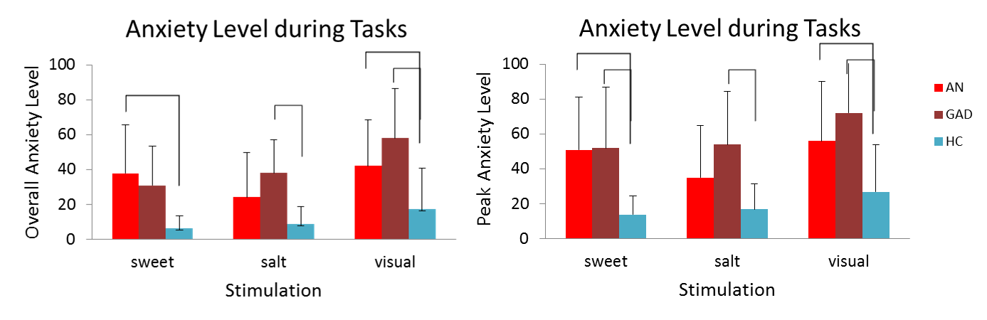
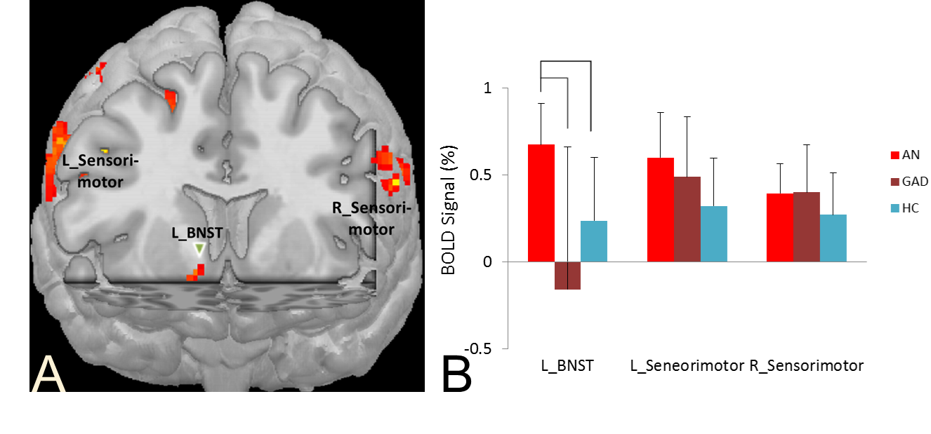
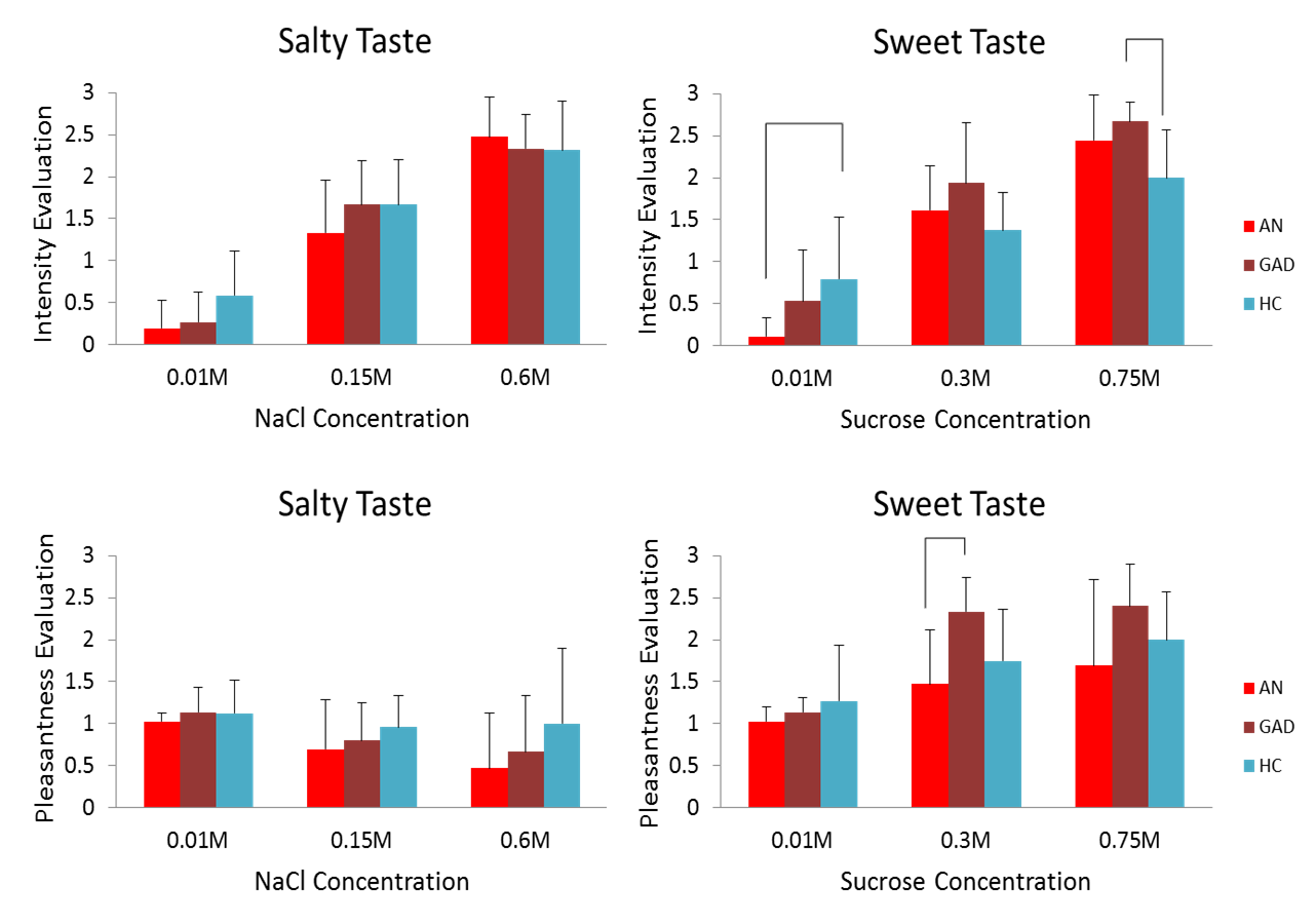
Most ANs and GADs had significantly high general anxiety level (Table 1). AN subjects had higher anxiety level than HCs during the milkshake and visual paradigm. In contrast, GAD subjects had significantly higher anxiety level than the HCs during all three paradigms (Figure 1). BNST in ANs was significantly activated during the milkshake paradigm, but not in the HCs and GADs (Figure 2). There were no significant differences in brain volumes (GM, WM, ICV) among groups (Table 2). No significant difference was present in BNST volume between ANs and HCs (Table 2). BNST was smaller in GADs than HCs in age-matched subgroups, however, not significant (ANCOVA with age as covariate) (Table 2). While there was no significant difference between ANs and HCs in the pleasantness ratings of sweet and salty tastes, AN subjects had lower sensitivity to the low-intensity sweet taste (Figure 3).Discussion & Conclusion
The milkshake stimulation triggered anxiety in AN subjects. Abnormal BNST function was discovered in AN subjects, which supports our hypothesis of increased BNST activity in AN related to food threat. AN subjects had lower sensitivity to sweet taste than HCs. This research opened a new line of inquiry into neuropathophysiology of AN, identifying new targets of treatment. A larger cohort study is warranted.Acknowledgements
This study was supported by the Klarman Family Foundation.References
1. Kaye WH, et al. Eur Eat Disord Rev. 2015;23(1):12-8. 2. Raney TJ, et al. Int J Eat Disord. 2008;41(4):326-32. 3. Kaye WH, et al. Am J Psychiatry. 2004;161(12):2215-21. 4. Godart NT, et al. Eur Psychiatry. 2000;15(1):38-45. 5. Walker DL, et al. Prog Neuropsychopharmacol Biol Psychiatry. 2009;33(8):1291-308. 6. Angeles-Castellanos M, et al. Neuroscience. 2007;144(1):344-55. 7. Wittmann W, McLennan IS. Horm Behav. 2013;64(4):605-10.Figures
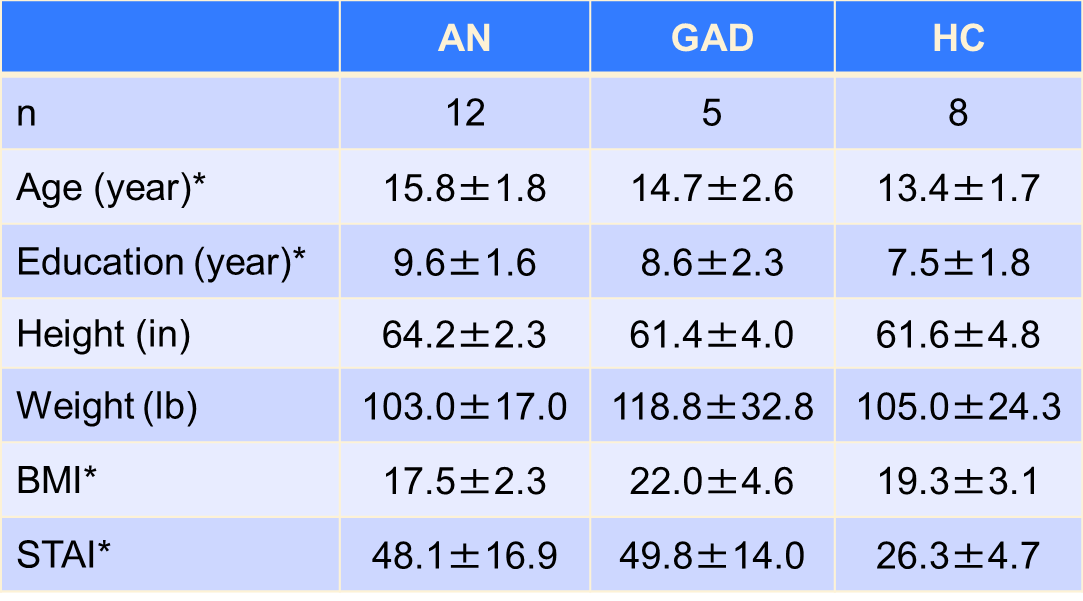
Table 1. Demographic & health information. STAI, state-trait anxiety inventory. *, ANOVA, significant difference between groups.