1965
Brain alternations after smoking cessation: An arterial spin labeling study1Department of Biomedical Imaging and Radiological Science, China Medical University, Taichung, Taiwan, 2Department of Radiology, China Medical University Hospital, Taichung, Taiwan
Synopsis
Although the unfavourable effects of cigarette smoking on the brain have been demonstrated in current smokers, it is unclear whether the neurotoxic effects of smoking on the brain are permanent or reversible after smoking cessation. Our results showed that ex-smokers had a decreased CBF when compared to never-smokers, especially in the posterior cingulate cortex (PCC). The present findings may explain in part the frequently reported cognitive dysfunctions in ex-smokers. However, the affected brain region was less extensive than the previous studies which compared current smokers and never smokers, suggesting the potential to partially recover from smoking-related CBF deficit.
Introduction
Chronic cigarette smoking is most common comorbidity worldwide, and is associated with the increased risk of pulmonary and vascular diseases. The focus of recent research has also shifted toward the understanding of the smoking-related brain diseases such as Alzheimer’s disease,1 as many neuroimaging studies have reported that cigarette smoking caused brain abnormalities including cortical thinning2 and regional brain atrophy.3 Although the unfavourable effects of cigarette smoking on the brain have been demonstrated in current smokers, it is unclear whether the neurotoxic effects of smoking on the brain are permanent or reversible after smoking cessation. Another less explored aspect in previous studies is that cerebral blood flow (CBF) has been considered as a powerful biomarker to explore neural dysfunction. However, till date, there is little information available regarding the relationship between CBF and smoking. Therefore, the central goal of this study is to evaluate the CBF using the noninvasive arterial spin labeling (ASL) technique for ex-smokers versus never-smokers. We also examined the dose-response relationship between smoking and CBF.Methods
Study design: There were 27 ex-smokers (8 males, 46.1 ± 13.3 years) and 83 never-smokers (27 males, 40.2 ± 14.8 years) with no history of smoking in this study. All volunteers had no history of cardiovascular diseases or psychiatric disorders according to self-completed questionnaires. Informed consent was obtained using IRB-approved protocol. MRI measurement: MRI scans were performed at a 3T Philips scanner and consisted of a T1-weighted magnetization-prepared rapid acquisition of gradient echo (T1-MPRAGE) and a pseudocontinuous arterial spin labeling (pCASL) sequences. The scan parameters of the T1-MPRAGE sequence were; TR/TE/TI = 8.1 ms/3.7 ms/1100 ms, and voxel size 1x1x1 mm3. Scan parameters of the pCASL sequence were; TR/TE = 4020 ms/14 ms, voxel size 3x3x5 mm3, labeling duration = 1.65s, post-labeling delay = 1.5 s, and 30 pairs of label and control images. Data analysis: ASL toolbox of MRICloud4 was used for CBF calculation. To test the association between smoking status category (ex-smokers and never-smoker) and CBF, the general linear model (GLM) correcting for sex, age, education year, and mini-mental state examination score was used. To assess the dose-dependent association, the second GLM correcting for above factors was performed in the ex-smokers group only, and the smoking status category term was replaced by a pack-years term.Results and Discussion
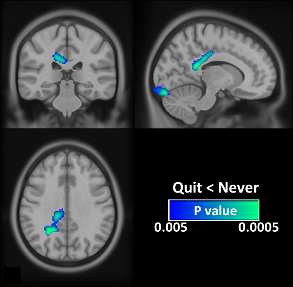
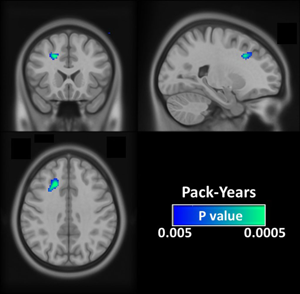
Associations between smoking status and CBF: Our results showed that ex-smokers had a decreased CBF when compared to never-smokers (Fig. 1). The most significant region of associations was in the posterior cingulate cortex (PCC). PCC is associated with cigarette, and alcohol, and illicit drug craving.5 Meanwhile, PCC forms a central node in the default mode network (DMN) of the brain, and DMN dysfunction is believed to be associated with poor cognitive function. The deficit CBF in PCC in ex-smokers found in this study may partly explain the greater decline in executive function in ex-smokers.6 Differences between ex-smokers and never-smokers were also found in the occipital lobe, which was known to have the higher density of nicotinic acetylcholine receptors.7 The neurotoxic effects of cigarette smoking can persist in some cerebral regions such as PCC and occipital lobe even after smoking cessation, but the affected brain regions were less extensive than previous studies which compared current smokers and never smokers.2, 3 In the light of the long-standing evidence of brain plasticity in adults, we speculate that this phenomenon may attribute to the potential reversibility of smoking-related CBF alteration in other cerebral regions. Dose-dependent association between smoking and CBF: In the ex-smokers group, a significant negative correlation between magnitude of lifetime exposure to tobacco smoke (pack-years) and CBF in the prefrontal cortex was observed (Fig. 2). Although the CBF decreases with the advanced age, findings here suggest that heavier cigarette consumption might accelerate this process and induce more pronounced cerebral perfection deficits.Conclusion
Using the ASL technique, our results showed that the brains of ex-smokers were functionally different from those of never smokers even after smoking cessation, especially in the PCC. The present findings may explain in part the frequently reported cognitive dysfunctions in ex-smokers. However, the affected brain region was less extensive than the previous studies which compared current smokers and never smokers, suggesting the potential to partially recover from smoking-related CBF deficit. This might serve as a useful motivation to encourage smoking quitting.Acknowledgements
No acknowledgement found.References
1. Barnes DE, Yaffe K. The projected effect of risk factor reduction on Alzheimer's disease prevalence. The Lancet. Neurology 2011; 10(9): 819-28.
2. Karama S, Ducharme S, Corley J, Chouinard-Decorte F, Starr JM, Wardlaw JM et al. Cigarette smoking and thinning of the brain's cortex. Molecular psychiatry 2015; 20(6): 778-85.
3. Brody AL, Mandelkern MA, Jarvik ME, Lee GS, Smith EC, Huang JC et al. Differences between smokers and nonsmokers in regional gray matter volumes and densities. Biological psychiatry 2004; 55(1): 77-84.
4. Li Y, Liu P, Li Y, Fan H, Su P, Peng SL et al. ASL-MRICloud: An online tool for the processing of ASL MRI data. NMR in biomedicine 2019; 32(2): e4051.
5. Bourque J, Mendrek A, Dinh-Williams L, Potvin S. Neural circuitry of impulsivity in a cigarette craving paradigm. Frontiers in psychiatry 2013; 4: 67.
6. Sabia S, Elbaz A, Dugravot A, Head J, Shipley M, Hagger-Johnson G et al. Impact of smoking on cognitive decline in early old age: the Whitehall II cohort study. Archives of general psychiatry 2012; 69(6): 627-35.
7. Jasinska AJ, Zorick T, Brody AL, Stein EA. Dual role of nicotine in addiction and cognition: a review of neuroimaging studies in humans. Neuropharmacology 2014; 84: 111-22.
Figures