1958
Quantitative MRI Volumetric Analysis of Rodent Brains as a Function of Age1Biomedical Engineering, University of Arizona, Tucson, AZ, United States, 2Psychology, University of Arizona, Tucson, AZ, United States
Synopsis
High resolution 3D-RARE magnetic resonance imaging (MRI) was carried out in the brain of rats (n=114) of varying ages and cognitive performance. Volumetric comparison of total intracranial volume (TIV) and total ventricular system (TVS) was performed using an atlas based analysis. The TIV increases from young adult to middle age but plateaus through old age. Average body weight increases from young adult to middle age but decreases from middle age to old age. No significant difference was found in TVS volume between ages or cognitive ability.
Introduction
Animal models play an important role in preclinical and translational studies of the human brain. MRI, being both non-invasive and inherently translational, can play an important role in comparing brain anatomy in animal models and humans, and atlas-based tools for neuroinformatics can be used to study age dependent changes in brain anatomy and function. The work herein presents initial results from a large cross-sectional study employing a rodent model of aging to investigate the neuroanatomical and epigenetic correlates of healthy cognitive aging. Initial analysis of MRI data has utilized a rat brain template and associated atlas (1) for comparison of rodent brains at ages ranging from young adult to middle age to old adult.Methods
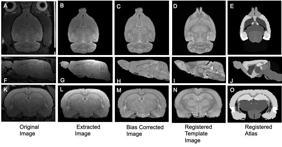
Male Fisher 344 rats (n=114) were acquired at young adult (6 months, n=48), middle aged (15 months, n= 38) and old adult (23 months, n= 28) ages. These animals underwent a battery of behavioral tasks over the course of 6 weeks resulting in each age group being sub-divided into 3 sub groups of high cognition, average cognition and low cognition using corrected integrated pathlength score which utilized last 12 spatial trails of the Morris water maze. At the end of the 6 weeks, body weights were measured and neurological MRI was carried out on a 7T Bruker Biospec (Bruker, Billerica, MA). A volume coil was used for excitation and a 4-channel phased array surface coil for reception. T2-weighted 3D Rapid Acquisition and Refocused Echoes (RARE) data, among other MRI scans, were collected with 150μm isotropic resolution, with the following imaging parameters: matrix size=256x192x128, TR=1500ms, ETL= 8, TEeff = 40ms, time of acquisition=76 min. Image Processing Raw images underwent brain extraction using a semi-automated process as well as bias correction due to non-uniform surface coil sensitivity using the ANTs software (2). A Waxholm Space Sprague Dawley T2-weighted template image (39 µm isotropic voxel) and labeled atlas (80 regions of the brain) (1), were resized to 150 µm isotropic voxels. The template image was registered to each individual animal in the study using linear and non-linear registration. The deformation fields produced from the registration were then applied to the labeled atlas and an in house Matlab code was used to calculate the volume of individual regions of interest (ROIs) in the brain. To compare ROI volumes across age and cognitive score, the volume of each ROI was normalized to total intracranial volume (TIV, 3).Results
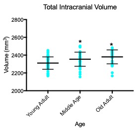
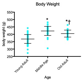
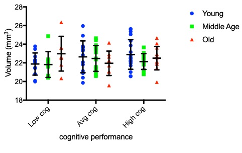
Example images demonstrating the processing steps are shown in Fig. 1. TIV calculated from semi-automatic brain extraction are plotted for each age group in Fig. 2 shows significant difference from young adult to middle age and young adult to old adult, however there is no significant difference between middle aged and old adult groups. Body weight (Figure 3) shows a significant increase from young adult to middle aged as well as old adult. However, there is a significant decrease from middle aged to old adult. The decrease in body weight from middle aged to old, while significant, was not substantial enough to bring the body weights back to the level of young adults. Total ventricular system volume is plotted for each age and cognitive group in Fig. 4.Discussion
For group-wise analysis considering different ages, total brain volume can be a confounding variable that needs to be accounted for (3, 4) The findings in this study indicate that rat brains are growing from young adult to middle aged and plateaus through old age. This challenges the assumption that the 6 month old age group can be considered a fully grown adult and it is possible that different regions of the brain are growing at different rates. Further study excluding the young adult population may yield interesting findings regarding the middle aged and old populations where the TIV growth has plateaued. Additionally, there was significant body weight increases from young adult to middle aged groups. This trend, however, did not persist as the bodyweight decreased significantly from middle aged to old adult. The total ventricular system was a region of particular interest because in human studies it has been shown to be an early indicator of aging and pathology. Because of its implications in humans, the growth of this region was of interest in characterizing rodent brain model constraints. No significant differences were observed in TVS volume throughout the age range or between cognitive performance. It is common in neuroscientific studies to study young animals in which the brain is not fully developed. While these studies can be informative, animal age must be considered as a factor that may influence results (4).Conclusion
Volumetric MR imaging detected differences in rodent total intracranial volume and the data suggests that the rat brains are continuing to develop past 6 months of age. Body weight measurements confirm the imaging findings to middle aged however there is a deviation at old age where TIV volume plateaus and body weight decreases significantly. These results will inform future analysis comparing regional brain volumes with age and cognition.Acknowledgements
No acknowledgement found.References
1. Papp EA, Leergaard TB, Calabrese E, et al. Waxholm Space Atlas of the Sprague Dawley Rat Brain. Neuroimage. 2014 Aug 15;97:374-86.
2. Avants BB, Yushkevich P, Pluta J. et al. The optimal template effect in hippocampus studies of diseased populations. Neuroimage. 2010 Feb 1;49(3):2457-66.
3. Barnes J, Ridgway GR, Bartlett J et al. Head size age and gender adjustment in MRI studies: a necessary nuisance? Neuroimage. 2010 Dec;53(4):1244-55.
4. McCutcheon JE and Marinelli M. Age matters Eur J Neurosci. 2009 Mar; 29(5): 997–1014.
Figures