1942
Neurite orientation dispersion and density imaging of rat brain microstructural changes due to middle cerebral artery occlusion at a 3T MRI
Zhenxiong Wang1,2, Wenzhen Zhu1, Shun Zhang1, Guiling Zhang1, Mehran Shaghaghi2, and Kejia Cai2
1Tongji Hospital, Tongji Medical College, Huazhong University of Science and Technology, Wuhan, China, 2Departments of Radiology, Department of Bioengineering, and the Center for MR Research, Chicago, IL, United States
1Tongji Hospital, Tongji Medical College, Huazhong University of Science and Technology, Wuhan, China, 2Departments of Radiology, Department of Bioengineering, and the Center for MR Research, Chicago, IL, United States
Synopsis
In this study we demonstrated the feasibility of longitudinal neurite orientation dispersion and density imaging (NODDI) in characterizing the microstructural alterations in rat brain tissues due to middle cerebral artery occlusion at a 3T MRI and validated the NODDI parameters with histology.
Introduction
Neurite orientation dispersion and density imaging (NODDI) can quantitatively evaluate specific microstructural changes in terms of neurite density and orientation distribution of axons and dendrites. There are no studies on NODDI of stroke animal models at clinical fields, a critical step for clinical translation given that MRI parameters are often field dependent. In the study, we attempt to demonstrate the feasibility of longitudinal NODDI in characterizing the microstructural alterations in rat brain tissues due to middle cerebral artery occlusion at a 3T MRI and to validate NODDI parameters with histology.Methods
All animal studies were approved by the institutional animal care and use committee. Wistar adult male rats (250 to 300 g, n = 11) were randomly selected as the infarction group. The suture was permanently fixed to the right internal carotid artery when it reached the base of right middle cerebral artery. As a comparison, 10 other rats (250 to 300 g) were included in the control group. For the control rats, skin incision was performed and carotid artery was separated from surrounding tissues with no ischemic procedure. After the animal preparations, a longitudinal multi-shell diffusion MRI protocol was performed for all rats at different time points of 0.5, 2, 6, 12, 24 and 72 h after operation respectively. Then, NODDI metrics of orientation dispersion index (ODI) and intracellular volume fraction (Vic) were generated using the publicly available NODDI toolbox and compared among groups at each time point. Meanwhile, the temporal evolution and the difference of hyperacute (including 0.5, 2 and 6h) and acute (including 12, 24 and 72 h) stroke periods for NODDI metrics were assessed. Following the last MRI, all rats were euthanized for histology staining of H&E, Nessile, myelin basic protein (MBP), and neurofilament (NF).Results
Infarction areas (LESION) showed significantly increased ODI and Vic compared to contralateral tissues (CONTROL) at all time points (P < 0.001, respectively) (Figure 1 and 2). There were no significant difference between CONTROL and healthy rat brain tissues on both left (“CONTROL_L”) and right (“CONTROL_R”) hemispheres for ODI and Vic. Lesion ODI increased gradually from 0.5 to 72 h, while its Vic showed a more complicated and fluctuated evolution with an initial reduction followed by an increase till 24 h (Figure 2). Moreover, ODI and Vic in lesions were significantly different between hyperacute (including 0.5, 2 and 6h) and acute (including 12, 24 and 72 h) stroke periods (P < 0.001, respectively), however, these metric values in contralateral brain tissues had no significant difference between different periods (Figure 3). Finally, histological findings validated the increased neurite orientation dispersion measured with ODI, increased intracellular volume measured with Vic and reduced neuron density in ischemic tissues by showing nerve fibers (neurites) bending, shrinkage, discontinuity, and thinning, neuron swelling, neurodegeneration (Figure 4).Discussion
In the present study, we demonstrated that the use of NODDI is feasible for the evaluation of rat brain microstructural changes due to MCAO at a 3T MRI. Lesion ODI increased gradually from 0.5 to 72 h, while its Vic showed a more complicated and fluctuated evolution with an initial reduction followed by an increase till 24 h. ODI and Vic were significantly different between hyperacute and acute stroke periods. Furthermore, NODDI metrics were confirmed by histological findings.Conclusion
NODDI metrics could reflect microstructural and pathological changes due to ischemic stroke in MCAO rats at 3T. This study helps to prepare NODDI for the diagnosis and management of ischemic stroke in translational research and clinical practice.Acknowledgements
This work was supported by grants from the National Natural Science Foundation of China (No. 81570462, No. 81730049 and No. 81801666), and partially supported by the NIH grants R21 EB023516 (Cai), R01 AG061114 (Tai&Cai), R21 AG053876 (Tai&Cai), the University of Illinois at Chicago Department of Radiology start-up funds (Cai). The authors thank Dong Kuang for his generous assistance with the histological analysis.References
No reference found.Figures
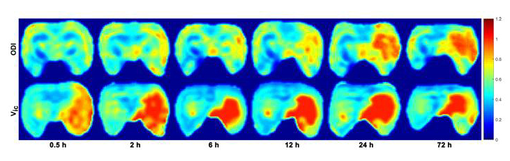
Figure 1. Representative NODDI metric maps at different
time points from a MCAO rat. The signal intensity of ODI and Vic maps in
the infarction areas is increased compared with contralateral brain tissues.
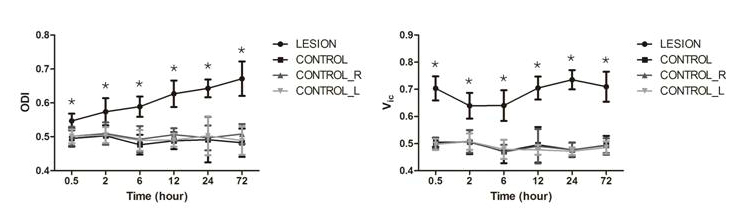
Figure 2. The temporal evolution of NODDI metrics from
different groups. * represents significant differences between LESION
and CONTROL.
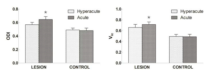
Figure 3. Comparisons of NODDI metrics in LESION and
CONTROL tissues between hyperacute and acute periods of ischemic stroke. *
represents significant difference between hyperacute and acute periods.
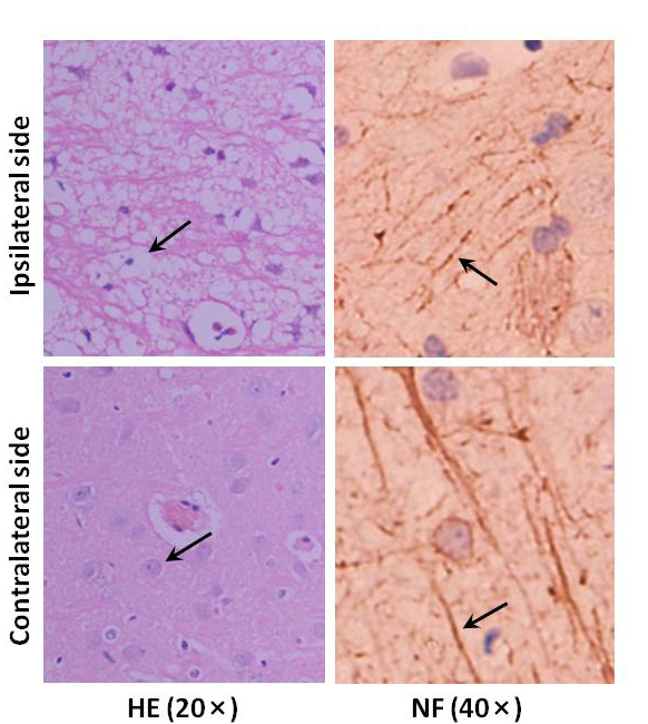
Figure 4. Representative histological staining of the ipsilateral
(LESION) and contralateral (CONTROL) sides at the time point of 72 h. Compared
with the contralateral brain tissue, H&E staining exhibits a large number
of abnormal neurons in morphology and structure, such as cellular swelling and
vacuolization, as well as nuclear degeneration; NF staining shows nerve fibers
bending, discontinuity and shrinkage. Black arrows exhibit the typical
histological changes in ipsilateral side and the normal structures in
contralateral side with different staining.