1937
Optogenetic modulation and rsfMRI mapping of depression related functional brain networks in the mouse
Laura-Adela Harsan1, Laetitia Degiorgis1, Julien Todeschi2, Lea Becker3, Maxence Thomas de la Pintière1, Victor Mathis3, Chrystelle Po1, and Ipek Yalcin3
1ICube: Engineering science, computer science and imaging laboratory, University of Strasbourg, Strasbourg, France, 2ICube: Engineering science, computer science and imaging laboratory, Neurosurgery Department, University Hospital Strasbourg, University of Strasbourg, Strasbourg, France, 3Institute de Neurosciences Cellulaires et Intégratives, University of Strasbourg-CNRS, Strasbourg, France
1ICube: Engineering science, computer science and imaging laboratory, University of Strasbourg, Strasbourg, France, 2ICube: Engineering science, computer science and imaging laboratory, Neurosurgery Department, University Hospital Strasbourg, University of Strasbourg, Strasbourg, France, 3Institute de Neurosciences Cellulaires et Intégratives, University of Strasbourg-CNRS, Strasbourg, France
Synopsis
The main objective of this study is to modulate and map the anterior cyngulate cortex (ACC) functional connectivity (FC) pathways underlying depression development in a mouse model. We use the optogenetic approaches to create the depression phenotype in mice, by activating the pyramidal ACC neurons expressing Channel rhodopsin 2 (ChR). We further use resting state functional MRI (rsfMRI) as non-invasive read-out of the effects at the level of functional brain connectivity. Four consecutive sessions of optogenetic ACC stimulation induced strong depression phenotype and major modifications of the functional mouse brain connectivity including perturbed mesocorticolimbic pathways and default mode network patterns
Introduction and objectives
Mood disorders, including depression, are increasingly seen as brain circuit pathologies rather than as region specific related disorders1. Recent data from the literature have identified the anterior cingulate cortex (ACC) as a key area of the affective component of chronic pain and a major player for further development of depression2. However, pathological perturbations of one brain area are rarely confined to a single locus; instead, they often spread to affect other regions and their connectional pathways. The main objective of this study is to modulate and map the ACC functional connectivity (FC) pathways underlying depression development in a mouse model. We use the optogenetic approaches to create the depression phenotype in mice3, by activating the pyramidal ACC neurons expressing Channel rhodopsin 2 (ChR). We further use resting state functional MRI (rsfMRI) as non-invasive read-out of the effects at the level of functional brain connectivity. Finally, we perform seed based analysis and graph theory approaches to identify the ACC functional circuitry signatures underlying depression.Materials and methods
Genetically modified mice expressing channelrhodopsin-2 and yellow fluorescent protein (Thy1-ChR2-YFP) in a subset of pyramidal neurons were used for optogenetic - rsfMRI studies. The animals (N=28) were subjected to: (i) surgery for glass fiber cannulas insertion along the whole vertical span of the ACC (1.7 mm long, cannulas MRI compatible, Doric Lenses) – allowing the light delivery via an optic fiber during optogenetic stimulation - (Fig. 1). (ii) a baseline rsfMRI brain scan, 1 week after cannula insertion; (iii) optogenetic stimulation for 30 minutes/day, during 4 consecutive days (repeated stimulations with 463 nm blue light) - inducing the anxio-depressive phenotype; (iv) behavioral evaluation of the depressive phenotype via standard “novelty suppressed feeding test”; followed immediately by (v) a second rsfMRI session to evaluate the functional connectivity features. 14 mice were subjected to “real” optogenetic stimulation and showed strong depression phenotype, while 14 mice (controls) underwent the same cannula implant procedures but the light was switched off during “dummy” stimulation sessions. Mouse brain rsfMRI was performed with a 7T animal scanner (Biospec 70/30, Bruker, Germany) and a combination of a transmit – receive volume coil (86mm) and a mouse brain adapted loop surface coil allowing the passage of the optogenetic cannulas (MRI, Bruker, Germany). The rsfMRI data was acquired under medetomidine anesthesia – initial bolus injection of 0.3 mg MD per kg body weight followed by continuous sc infusion of MD at 0.3 mg per kg body weight per hour. A one-shot GE-EPI sequence (TE / TR = 15 ms / 2000 ms; resolution = 0.14 × 0.21× 0.5 mm³; 500 volumes of 31 axial slices) was used for rsfMRI, This data was spatially normalized on the Allen Mouse Brain Atlas and frequency filtered (<0.01Hz).Results and Discussion
Four consecutive sessions of optogenetic ACC stimulation induced strong depression phenotype and major modifications of the functional mouse brain connectivity. Fig 1 exemplifies the connectivity of dorsal ACC, before (A) and after (B) optogenetic stimulations. Overall decrease of FC is noticed between ACC and the rest of the cortical areas, notably prefrontal areas (PFC) (red – positive correlations) but also with subcortical nuclei, such as Nucleus Accumbens – NAc. Both PFC and NAc are known hubs of the mesocorticolimbic circuitry, and their perturbed patterns of connectivity in depression suggest altered cognitive processing of motivation, aversion and reward. Additionally, our data indicate a major impact of optogenetic stimulation on the default mode network (DMN) pattern and on its cross-talk with the rest of the brain. Modifications of the default-mode network (DMN) is one of the most widely replicated neuroimaging findings in major depressive disorder (MDD) in humans4. Moreover, human fMRI studies demonstrate that mood disorders disrupt the relationship between DMN and the other networks of the brain, similar to our observations in mice.Acknowledgements
No acknowledgement found.References
1Helm et al. Neuropsychiatr Dis Treat. 2018 Oct 17;14:2715-2737; 2Rolls et al., Cereb Cortex. 2018 Nov 12. doi: 10.1093/cercor/bhy236; 3Barthas et al., Biol Psychiatry, 2015, 77:236-245.; 4Brakowski et al., J Psychiatr Res. 2017 Sep;92:147-159.Figures
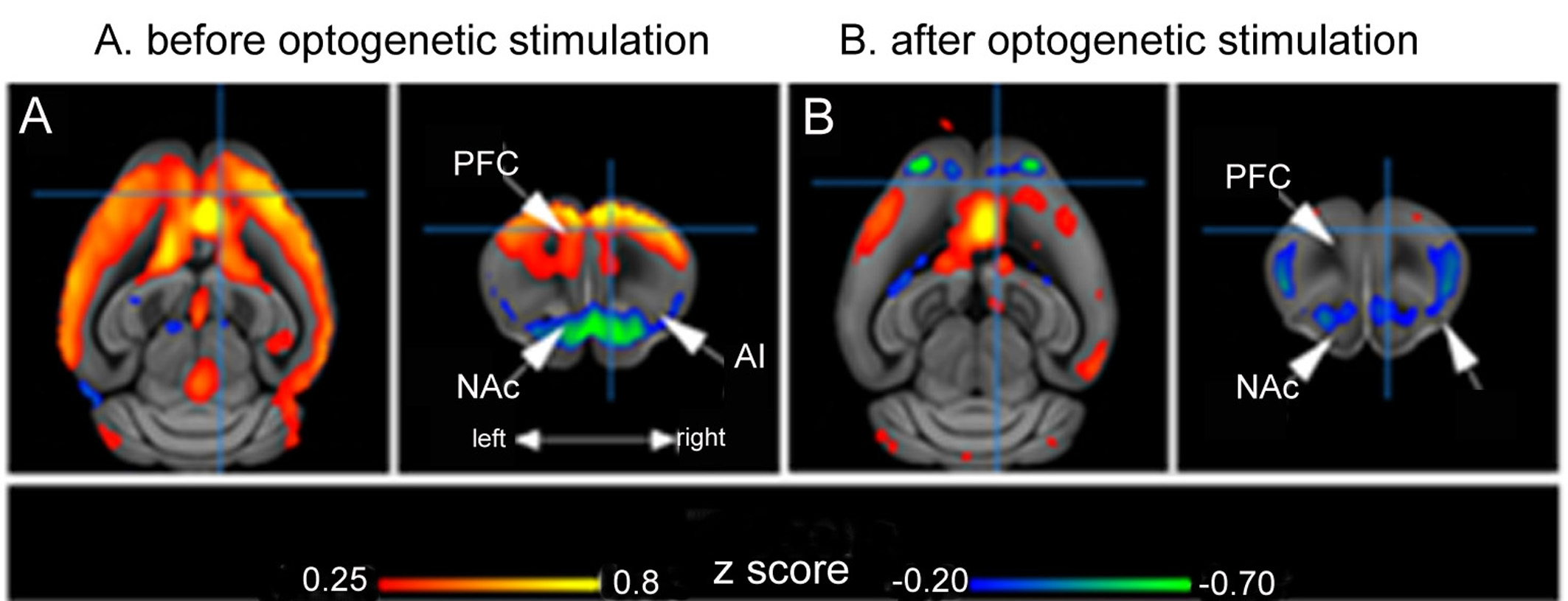
BOLD rs-fMRI mean correlation map depicting FC patterns of dorsal ACC (A) before and (B) after depression induction via optogenetic stimulation. The color scale indicates the correlation values (positive correlations: dark red to yellow; and negative correlations: blue to green).