1928
Imaging Changes in Blood Brain Barrier Permeability Following Repetitive Mild Traumatic Brain Injury.1Psychology, Northeastern University, Boston, MA, United States, 2A.A. Martinos Center for Biomedical Imaging, Massachusetts General Hospital, Charlestown, MA, United States
Synopsis
Failure in the blood brain barrier (BBB) lies at the foundation of small vessel disease that is precursor to neurodegenerative diseases. There are multiple risk factors leading to increased permeability in the BBB; one prevalent risk factor is repetitive mild TBI. Imaging the subtle changes in BBB permeability is not possible with standard imaging protocols. With quantitative ultra-short time-to-echo, contrast-enhanced (QUTE-CE) MRI, using the superparamagnetic iron oxide nanoparticle ferumoxytol, we can image changes in BBB permeability following rmTBI. With this novel methodology we can measure the immediate effects of rmTBI on changes in BBB permeability across the entire rat brain.
Introduction
TBI is one of the most prevalent risks of death and disability in young people, with about 1.6 million reported per year in the US. Mild-TBI (mTBI) is characterized as a negligible loss of consciousness with minimal neuropathology; and is estimated to account for 70-90% of all TBI cases. Micro-vessel disruption plays substantial role in primary, secondary and chronic effects of TBI, and initiates a process that induces molecular, biochemical, and cellular changes, which in turn contribute to BBB disruption that leads to neuronal damage and death over time. Here we introduce new method for quantifying BBB disruption by introduction of intravascular superparamagnetic iron oxide nanoparticle (SPION) contrast agent (CA), Ferumoxytol (AMAG Pharmarceuticals, Waltham, MA), and the measurement of CA leakage into the surrounding parenchyma without evidence of capillary rupture.Methods
Sprague dawley male adult rats, weighed approximately 400gm, were divided into three groups. Control group with no hit, TBI group with one-hit (n=9) and TBI with three-hit (n=7) which received 3 mild hits for 3 consecutive days. Quantitative Ultra short Time to Echo – contrast enhanced (QUTE-CE) MRI data was acquired on all rats before and immediately after ferumoxytol administration (for a 200pg/ml iron concentration in the blood) every 7 minutes for one and half hour. Data was registered to MRI Rat brain atlas (Ekam Solutions LLC, Boston MA) and segmented into 174 ROIs. Quantitative global percentage change in signal for each ROI (174 ROIs) compared to baseline (pre-contrast imaging) was computed for each group. TBI was introduced using momentum exchange model (et al Vieno), and we reproduced consistently the 7.4, 9.3 and 11.2 m/s impact velocities described for mild, medium and severe rat head injury, respectively. The data reported here all came from the 7 m/s impact velocities as determined using accelerometer. QUTE-CE MRI images were obtained using a Bruker Biospec 7T/20 cm USR horizontal magnet (Bruker, Massachusetts, USA) with ParaVision 6.0.1 acquisition software. UTE images were acquired with TE=0.013 ms, TR= 4 ms, FA= 20°, RF bandwidth 200kHz, field of view (FOV) 3×3×3 cm3, matrix 180×180×180. To align the acquired UTE images with MRI rat brain atlas, anatomical T2-weighted RARE images were taken with the parameters: TE 7 ms, TR 3000 ms, RARE factor 4, FA 90°, averages 3, FOV 3×3 cm2, matrix 180×180, slice thickness 5 mm, number of slices 64.Results
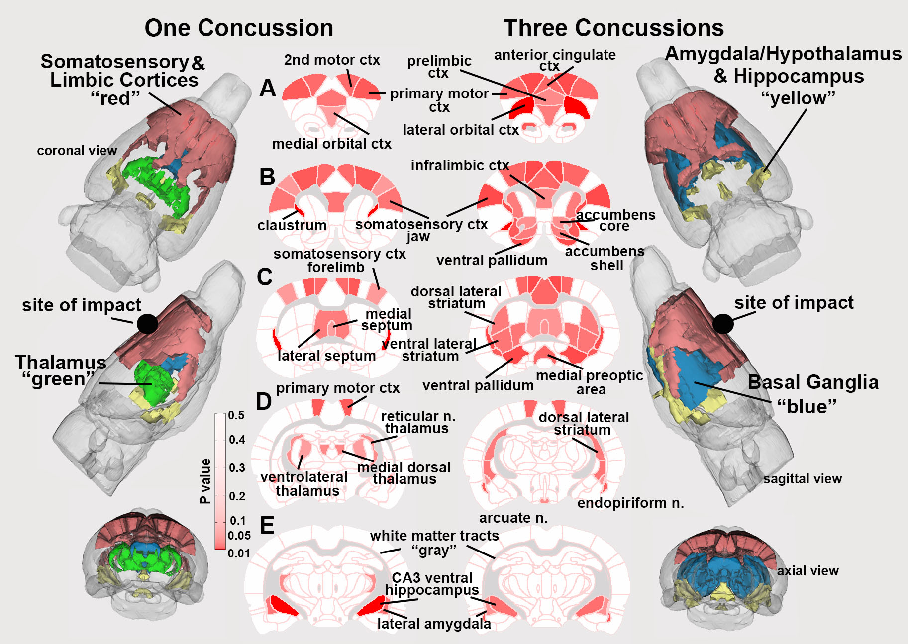
The site of impact was around the rostral cranium, directly affecting the underlying motor cortex. As shown in figure 1, the primary motor cortex and secondary motor cortex regions demonstrated significant increase of ferumoxytol accumulation following three mild concussions. Impacts on the same site repetitively within short time intervals may have produced a unique mechanical impact leading to the significant change in cortex regions and striatum regions. Other regions significantly affected are hypothalamus, basal ganglia, cerebellum and brainstem.Discussion
With a signal mTBI, the BBB barely changed in rat brain which is consistent with other findings, minor alterations the BBB integrity and little structural change. While with three concussions, BBB damage occurred with ferumoxytol leakage from blood vessels in several regions including cortex, striatum, hypothalamus, thalamus, and brainstem. This was confirmed with several human and animal studies reporting that repetitive mild TBI separated by short intervals pose a greater risk than single TBI and cortex, striatum and cerebellum are particularly vulnerable to mTBI. There are some limitations of our study we followed scanning the rats for 70 minutes after contrast agent administration. As shown in the result part (figure 2), around 70 minutes, signal intensity still increased which means the peak time of ferumoxytol leakage and accumulation is beyond 70 minutes. Future work will add more scans after 70 minutes to get the peak time and the ferumoxytol leakage curve.Summery
Rats underwent 1 hit show small difference in ferumoxytol accumulation, indicating blood brain barrier was not disrupted. Rats underwent 3 repeated hits showed significant difference in ferumoxytol accumulation in extravascular space indicating severe BBB disruption.Acknowledgements
No acknowledgement found.References
Kulkarni P, Morrison T, Ferris C. et al. Neuroradiological Changes Following Single or Repetitive Mild TBI. Front. Syst. Neurosci. 13:34. doi: 10.3389/fnsys.2019.00034.
Gharagouzloo C, Timms 2, Qiao J et. al. Quantitative vascular neuroimaging of the rat brain using superparamagnetic nanoparticles: New insights on vascular organization and brain function. Neuroimage. 2017 Dec;163:24-33
Figures