1900
Application of GAN in optimizing compressed sensed MR imaging of brachxial plexus
Yan Fu1, Tianjing Zhang2, Haixia Li3, Yichen Tong3, Xiangchuang Kong4, and Dingxi Liu4
1EPFL, Lausanne, Switzerland, 2Philips Healthcare, Guangzhou, China, 3Sun Yat-Sen University, Guangzhou, China, 4Radiology, Union Hospital,Tongji Medical College, Huazhong University of Science and Technology, Wuhan, China
1EPFL, Lausanne, Switzerland, 2Philips Healthcare, Guangzhou, China, 3Sun Yat-Sen University, Guangzhou, China, 4Radiology, Union Hospital,Tongji Medical College, Huazhong University of Science and Technology, Wuhan, China
Synopsis
In routine MRI clinical application of compressed sensing, the image quality is often not useful for diagnosis when its CS(Compressed SENSE) acceleration factor is beyond a certain level(e.g. 8 or 10). It is desirable to further accelerate MR sequences in multiple applications such as brachial plexus nerve, coronary artery, and so on. It is possible to use generative adversarial network(GAN) models to further optimize the imaging workflow by improving the image quality of data acquired with high CS factors.
Introduction
Generative adversarial network (GAN) is now broadly applied in medical imaging [1]. Compressed sensing has attracted significant attention in medical imaging, which reduces the acquisition time[2]. However, high acceleration factor of compressed sensing would meanwhile compromise the signal-to-noise ratio, affecting image quality and diagnostic performance. In the clinic,3D contrast-enhanced nerve-view imaging provides high diagnostic value for brachial plexus nerve trauma, tumor, and so on.However, a relatively long acquisition time (above 10 minutes) limits its clinical application. In a previous study, we have evaluated the image quality and capability of diagnosis of accelerated 3D Nerve-view sequence, the images of CS factor above 8 has no diagnosis value. The SENSE = 2 and CS factor = 4 - 20 images are well collected as data pairs, which is very suitable for generative adversarial network experiment design and training. Accordingly, we designed a study to validate the feasibility of GAN models in optimizing MR images generated from CS factor = 10.Material and Methods
In a consecutive cohort of 35 patients with suspected disease of brachial plexus underwent MR studies. 3D Nerve-VIEW sequences with 4 CS accelerating factors(4,6,8,10) and a traditional 3D Nerve-view with 2-fold parallel imaging(SENSE) as a clinical reference were obtained on a 3.0T scanner(Ingenia CX, Best, Philips Healthcare), 100 slices of different acceleration factors were acquired for all patients. Images were graded by two experienced radiologists in MR neurography. The results showed that images of CS factor =10 have no diagnosis value. However, those images with CS factor = 10 still have basic structures of brachial nerve whose image quality. Moreover, the slice position, slice number is the same for different acceleration factors in each patient’s image acquisition, which could be naturally used for the experiment design in a generative adversarial network. Accordingly, we designed an experiment to optimize the image quality of CS factor = 10. From all the acquired images, images acquired with SENSE factor = 2 are the best, so we use those images as ground truth. In our study, we used images with SENSE=2 as a discriminator, images with CS factor = 10 as a generator. A mainstream generative adversarial model: pix2pixHD is applied in our study. This model is typically used for high-resolution image-to-image translation[3]. Original image size is 1152*1152 in Dicom format, and we randomly crop the image into 1024*1024 then resized it into 512*512. Training epoch is 50, batch size 1 with initial learning rate 2e-4 for Adam optimizer. The parameter for the exponential decay rate for the 1st-moment estimates is 0.5 and for the 2nd-moment estimates is 0.999. The networks are implemented in the TensorFlow and Pytorch frameworks on an NVIDIA GEFORCE RTX 1080 Ti GPU.Results
Peak signal-to-noise ratio(PSNR) and structural similarity index(SSIM) was chosen to perform image quality evalution[5]. The quantitative results for the two models are presented in Table 1. As observed, relative to CS = 10 images, pix2pixHD model has an obvious improvement in both PSNR and SSIM indexes. We also performed a blind reader study on the images generated from 10 test patients. Two experienced radiologists with 10 years of experience scored the images generated from the test samples by the three methods in terms of image quality on a point scale (1=unacceptable, 5 = best). The scoring results are shown in Table 2. It shows that the scoring for Pix2pixHD model is better than CS = 10.To show the image enhancement effects of the pix2pixHD networks, we took representative slices, as shown in Fig. 1Discussion
In this study, we mainly validate the performance of pix2pixHD model in brachial plexus MR imaging. Our focus is on the improvement of image details. MR images in brachial nerves with CS factor=10 could not clearly demonstrate the structures of the brachial plexus and its surrounding tissue. Moreover, the lesion display is not very clear either. The main reason we choose pix2pixHD model for image enhancement and denoising is that this GAN model is typically used for high-resolution image-to-image translation. Its primary advantage include multi-scale generator and discriminator for high-resolution image generation; multiple editable outputs could be generated with different input conditions. In our task, the high-resolution image generation could enable us to figure out more lesion details. In future, we could potentially design a cascaded GAN model for further denoising of the generated images. From two radiologists' perspective, our generated images enhance the signals of brachial nerves. However, there is some extra noise included in our generated images as well. More algorithm improvement and multicenter clinical validation are needed before the generated images from CS = 10 can be applied in real clinical applications.Conclusion
In conclusion, this study innovatively applies an advanced generative adversarial network in brachial nerve MRI. Such a method effectively improves both the image index and subjective scoring in comparison with CS = 10 images; it also enhances the demonstration of lesion details and nerve structures of CS = 10 images. In future, more denoising approach should be applied to make the algorithm’s performance more satisfactory for clinical diagnosis.Acknowledgements
No acknowledgement found.References
- X. Yi, E. Walia, and P. Babyn, “Generative adversarial network in medical imaging: A review,” 2018.
- Geethanath S1, Reddy R2, Konar AS3, Imam S2, Sundaresan R4, D R RB3, Venkatesan R4."Compressed sensing MRI: a review." Crit Rev Biomed Eng. 2013;41(3):183-204.
- Ting-Chun Wang, Ming-Yu Liu, Jun-Yan Zhu, Andrew Tao, Jan Kautz1, Bryan Catanzaro,"High-Resolution Image Synthesis and Semantic Manipulation with Conditional GANs arXiv:1711.11585v2 [cs.CV] 20 Aug 2018
Figures
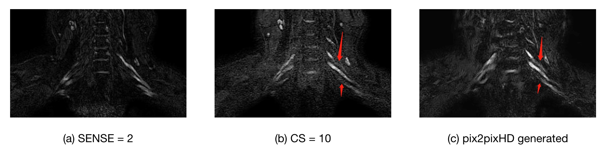
Figure 1. Example
pictures of SENSE = 2, CS = 10 and images generated from pix2pixHD
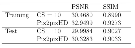
Table1. Quantitative
measurement results of pix2pixHD model
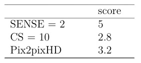
Table2. Subjective scoring
results from two experienced radiologists