1897
Deep-Learning Reconstruction Method for Improved High-Resolution Imaging of the Lateral Rectus-Superior Rectus Band in a Clinical Setting1GE Healthcare, New York, NY, United States, 2Radiology, Columbia University, New York, NY, United States, 3GE Healthcare, Calgary, AB, Canada
Synopsis
The purpose of this study was to determine the effectiveness of using deep learning reconstruction (DL Recon) for improved signal-to-noise (SNR) allowing for higher resolution imaging of the lateral rectus-superior rectus band of the orbit without substantial increase in scan/exam time. Evaluation of this new reconstruction technique was performed on a group of 5 volunteers with results indicating that higher resolution protocols can produce images with increased SNR and removal of noise leading to higher confidence in identifying the lateral rectus-superior rectus band.
Purpose
Rectus muscles are responsible for globe motion in the orbit. Collagenous rings within the Tenon’s fascia encircle the rectus muscles near the globe equator and are also termed rectus pulley system. The pulleys are themselves interconnected by suspensory connective tissue that extend between each adjacent rectus pulley system. One such connective tissue is called the Lateral Rectus Superior Rectus band (LR- SR band). Degeneration of this band has been implicated in the development of 2 related forms of strabismus: heavy eye syndrome and sagging eye syndrome. Both LR-SR band discontinuity (and rupture) and supratemporal displacement have been reported as features of band degeneration in patients with strabismus. Improved visualization of this structure would aid in early diagnosis of these entities. The aim of our study is to test a new deep learning reconstruction algorithm to improve detection of this important orbital structure.Methods
The study was performed on 3T 70cm bore MR scanners (Discovery MR750w, GE Healthcare, USA) using GE Signa MRI Brain Array Coil (8 channels, High Resolution). The data collected was FSE T2 anatomic scan in the qusi-coronal plane with the following parameters: 12cm FOV, 2mm slice thickness, 3800 millisecond TR, 82 millisecond TE, 41 kHz bandwidth and a matrix of 300 x 300, 18 slices with a total scan time of 3:12 per scan. Once data was acquired using conventional reconstruction, a second data set was reconstructed off-line with the DLRecon method. The DLRecon method comprised a deep convolutional residual encoder network trained from a database of over 10,000 images to reconstruct images with high signal-to-noise ratio and high spatial resolution. The network offered a tunable noise reduction factor to accommodate user preference. The DLRecon network was embedded into a new reconstruction pathway that operated in parallel with the conventional reconstruction such that two sets of image series were generated from a single set of raw MR data. All deep learning reconstruction sequences used a 75% noise reduction calculation. Imaging parameters for this imaging data set described above were developed by the author to produce resolution/voxel size that would greatly improve visualization these above-mentioned anatomical structures. The reviewer(neuroradiologist) subjectively assessed the quality and detail of the images by comparing the two reconstruction types with respect to visibility of the LR-SR band.Results
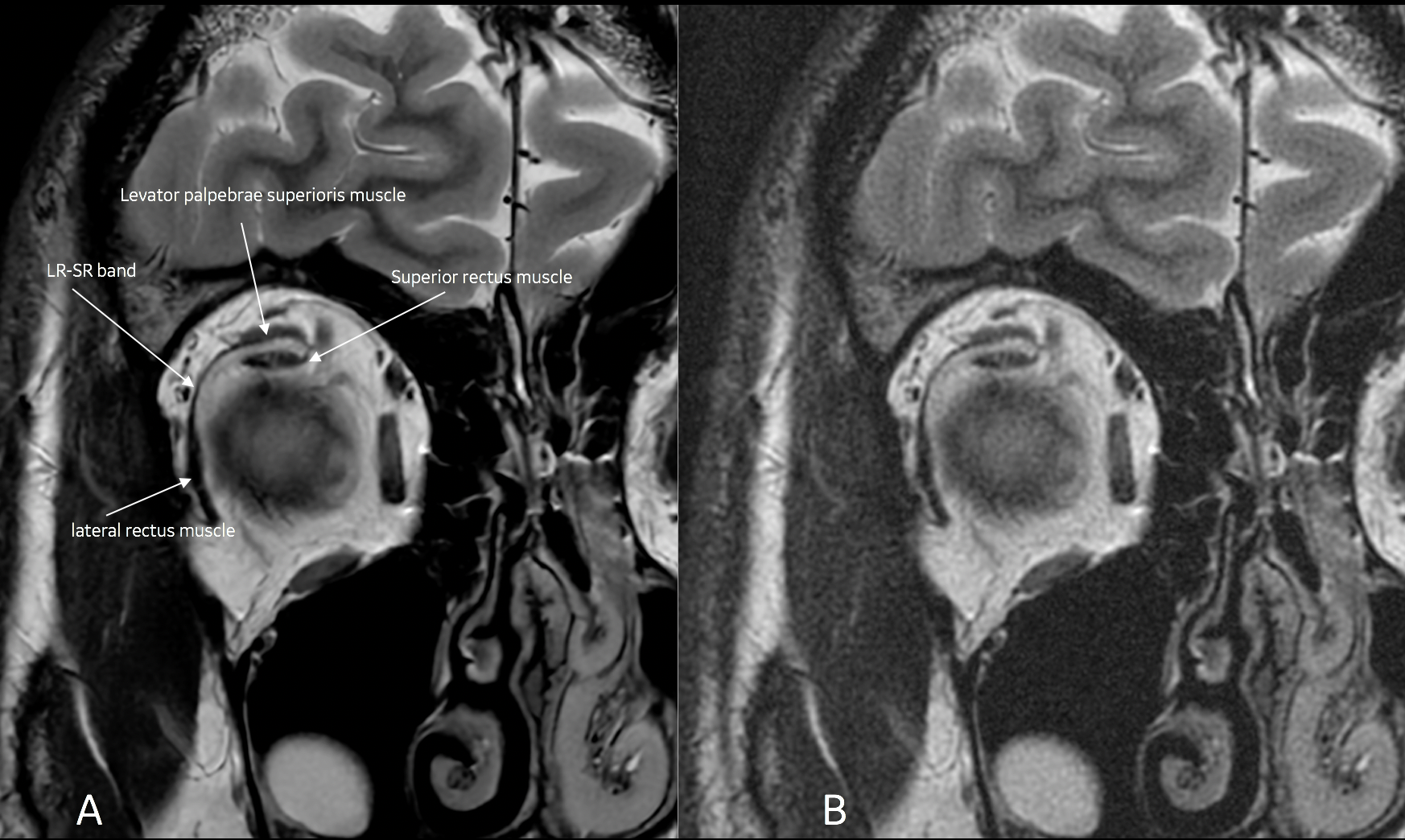
There was subjective improved visualization of the LR-SR band on the DL reconstruction algorithm compared to non DL algorithm, Furthermore there was improved distinction between the levator palpebrae superioris muscle and the superior rectus muscle on the DL reconstruction algorithm (Fig 1).Discussion
Our results provide preliminary DLRecon image data for improved visualization of the LR-SR band without minimal scan time increases. Increasing resolution (voxel size) generally will improve visualization for anatomical structures, such as those listed in this study, when properly balanced with suitable signal-to-noise to provide an image free as possible of noise. This proper balance of SNR typically comes at the expense of increase scan time, SAR and likelihood of patient motion (due to longer scan). SNR calculations taken from identical slice locations between the two reconstruction methods shows substantial 62% increase in signal in the orbital globe with the DL Recon image (Fig 2). This significant increase in SNR is key in allowing for this type of high-resolution protocol for imaging these structures minus the traditional methods to achieve the same quality of scan. The result from this data shows DL approach to noise reduction can allow for higher resolution with very minimal increase to scan time to produce images free of noise, increased SNR and excellent structure detail in the orbit (Fig 3.).Conclusion
In this work, we demonstrated that deep learning reconstruction method can provide significant improvement to SNR through noise reduction allowing users to modify current protocols to increase resolution for enhanced delineation of the lateral and superior rectus band. This newly developed approach to noise reduction allows the potential user to greatly improve current clinical protocols for many of their exams, but also gives them the flexibility to strive for higher resolution for specialized protocols/exams without degradation to of image quality and/or substantial scan time increase.Acknowledgements
No acknowledgementsReferences
1. S.H. Patel, M.E. Cunnane, A.F. Juliano, M.G. Vangel, M.A. Kazlas and G. Moonis. Imaging Appearance of the Lateral Rectus-Superior Rectus Band in 100 Consecutive Patients without Strabismus. American Journal of Neuroradiology September 2014, 35 (9) 1830-1835; DOI: https://doi.org/10.3174/ajnr.A3943C
2. Chaudhuri Z, Demer JL. Sagging eye syndrome: connective tissue involution as a cause of horizontal and vertical strabismus in older patients. JAMA Ophthalmol 2013;131:619–25
Figures