1892
Differentiation of adult supratentorial ependymomas from high grade gliomas using textural features based on diffusion weighted imaging.1Neuroimaging and Interventional Radiology, NIMHANS, Bengaluru, India, 2Symbiosis Centre for Medical Image Analysis (SCMIA), Symbiosis International University, Pune, India
Synopsis
High grade extraventricular supratentorial ependymoma’s in adults are uncommon neoplasms with imaging features that can mimic cortical tumors if small and high-grade gliomas (HGG) if large. No previous work has tried to discriminate ependymoma from high grade gliomas using MRI. Our work evaluates preoperative diffusion weighted imaging for discrimination of ependymomas from grade III and grade IV gliomas using textural analysis. Results demonstrate significant differences in the histogram and first order textural features derived from diffusion weighted imaging in cases of ependymomas and high-grade gliomas.
INTRODUCTION
Supra-tentorial extraventricular ependymomas constitute a rare form of adult intracranial tumors and show imaging features similar to those of cortical tumors and high-grade gliomas namely anaplastic astrocytomas or glioblastomas [1, 2]. This is especially true for the cerebral extra-ventricular ependymomas as the location of the neoplasm does not support the diagnosis because of their relative rarity. Advanced MRI methods like Diffusion weighted imaging (DWI), perfusion MRI and MRS shows imaging features of ependymoma simulate malignant gliomas like restricted diffusion within the solid tumor and elevated choline on MRS indicating high cellularity and increased CBV on perfusion MRI suggestive of neoangiogenesis. All these features make discrimination of ependymoma difficult from high grade gliomas. [3, 4]. Treatment strategy and prognosis of ependymomas differs significantly and a standard course of management including radiation and chemotherapy in case of ependymomas is unclear [2, 5]. It is therefore important to predict the tumor type preoperatively using imaging, thus helping in prognostication and optimizing the treatment planning and efficacy. This study aims to discriminate supratentorial ependymomas using an underlying radiomics signature derived from diffusion weighted imaging from high grade gliomas.METHOD
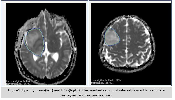
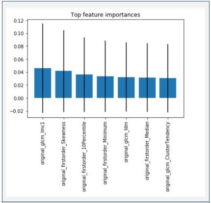
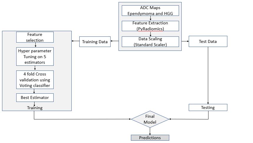
This retrospective study included 17 Ependymomas (age 21±11 years, 8 Females) and 41 HGG [21(grade-III and 20 grade-IV), (age 45±13 years, 18 Females)] patients, classified with tumor histopathological characterization done using WHO 2016 classification. MRI data included diffusion weighted MRI performed on 3T MR scanner (Philips Achieva) using a 15-channel head coil, 3T MRI (Siemens Skyra) and 1.5T (Siemens Aera) using 20 channel and 8 channel coils respectively. Data was acquired with TR 3000 ms, TE 73 ms, Voxel Size 2x2x4, b-values 0 and 1000 on 3T Philips scanner and TR 3800 ms, TE 83 ms, Voxel Size 2x2x5, b-values 0 and 1000 for Siemens scanner. Image processing and quantitative analysis of MRI data were performed using 3D Slicer, PyRadiomics and Voting Classifier from SKLEARN [6].Tumor masks generation and intensity thresholding was performed on ADC maps of HGG and ependymoma using segmentation wizard extension of 3D slicer version 4.10.2 (Figure1). Segmentation was cross validated by an experienced neuroradiologist. Total 42 features (18 first order features and 24 GLCM features) were extracted using PyRadiomics open source python package after z-score normalization of the original ADC maps. The obtained data was scaled using StandardScaler. Scaled data was divided into training set (52 training examples) and holdout set (6 examples). Feature importance was calculated on the training dataset using ExtraTreeClassifier from sklearn (Figure2). Principal component analysis (PCA) was used for reducing the dimensions of the training data. Following five estimators were trained- Support Vector Machine, Gradient Boosting Classifier, XGBoost Classifier, Logistic Regression and Bagging Classifier. Hyper parameters of these estimators were tuned using four-fold Grid Search CV. These estimators with tuned parameters were combined using a voting classifier. Four- fold Cross validation was done using cross_validate module of sklearn. Best estimator returned from cross validate instance was used as the final model (Figure3).
RESULTS
69.23% cross validation accuracy and 0.573 cross validation ROC AUC score were obtained. Final model prediction on holdout dataset yielded 83.33% accuracy with 0.166 mean squared error and 0.777 F1 score. The top ranked textural features for discrimination included – skewness, 10th Percentile, Minimum, Median, Cluster Tendency, glcm-idm and glcm-imc1. Since the dataset was small and number of features were comparable to the size of the data some kind of dimensionality reduction technique was needed to avoid overfitting on the data. Even after reducing dimensionality the performance of individual estimator was not satisfactory so Voting classifier was used to combine the predictions from individual (Weak Classifiers) to obtain a strong classifier.DISCUSSION
In the current study we investigated textural features of supratentorial extraventricular ependymomas and compared them with high grade gliomas. Supratentorial ependymomas are rare neoplasms and only a few case series have been described in the literature. As these tumors resemble high grade neoplasms and their management strategies are unclear it is clinically relevant to discriminate them from high grade gliomas. In the current study we found textural features derived from DWI images useful for discriminating them from high grade gliomas. Our diffusion MRI based image classifier was able to discriminate these rare neoplasms from commonly occurring high grade gliomas. Important limitation of the current study is small number of subjects however due to relative rarity of supratentorial ependymomas it is very difficult to accumulate large numbers of these neoplasms. It is important to incorporate the imaging features of these neoplasms in the image classifier algorithms as they do not have characteristic imaging features and are histologically and prognostically distinct entity.CONCLUSION
High grade supratentorial ependymoma’s in adults can be differentiated from high grade gliomas using textural features and machine learning algorithm can be used to classify HGG and ependymoma with decent accuracy.Acknowledgements
No acknowledgement found.References
1. Leng, X., et al., Magnetic resonance imaging findings of extraventricular anaplastic ependymoma: A report of 11 cases. Oncol Lett, 2016. 12(3): p. 2048-2054.
2. Byun, J., et al., Supratentorial Extraventricular Ependymoma: Retrospective Analysis of 15 Patients at a Single Institution. World Neurosurg, 2018. 118: p. e1-e9.
3. Wu, J., T.S. Armstrong, and M.R. Gilbert, Biology and management of ependymomas. Neuro Oncol, 2016. 18(7): p. 902-13.
4. Shintaku, M. and K. Hashimoto, Anaplastic ependymoma simulating glioblastoma in the cerebrum of an adult. Brain Tumor Pathol, 2012. 29(1): p. 31-6.
5. Ruda, R., et al., EANO guidelines for the diagnosis and treatment of ependymal tumors. Neuro Oncol, 2018. 20(4): p. 445-456.
6. Pedregosa, F., et al., Scikit-learn: Machine Learning in Python. Journal of Machine Learning Research, 2011. 2011(12): p. 2825-2830.
Figures