1884
Regional gray matter volume predicts HIV-positive patients with HAND using a machine learning approach
Danhui Fu1, Guanqiao Jin1, Lidong Liu1, Wenjuan Deng1, Wei Zhang1, Qianlin Ding1, Weiyin Vivian Liu2, Long Qian2, and Danke Su1
1GuangXi Medical University Cancer Hospital, Nanning, China, 2MR Research, GE Healthcare, Beijin, China
1GuangXi Medical University Cancer Hospital, Nanning, China, 2MR Research, GE Healthcare, Beijin, China
Synopsis
Conventional neuropsychometric tests could not early detect HIV-associated neurocognitive disorder (HAND) on HIV-patients. More objective indicators with neuroimaging techniques have offered to sub-classify patients with specific symptoms. Significant differences of regional gray matter volumes (rGMV) betweenHIV-positive group and age-matched healthy group was found in our preliminary study. Ten major rGMV well distinguished HIV-positive patients with HAND from those without HAND. Our study showed rGMV might be an indicator to explain for neurocognitive impairments.
Introduction
Abnormal brain structure of HIV carriers occurs before HIV-positive patients receive treatments of reverse virus transcription1. Severity of HIV may affect substantial reconstructionof default mode network (DMN), task engagement and perception-related network connectivity2. Moreover, neuroimaging techniques may offer more objective scientific evidences for future diagnosis3. Thus, this study aims to explore the role of gray matter volume (GMV) in determining HIV-positive patients from normal healthy volunteerswitha machine learning approach.Materials and Methods
27 HIV-positive patients (14 males and 13 females) without anti-virus therapy and 14 healthy volunteers (8 males and 6 females) were enrolled in our study. 18 HIV-positive patients with HAND and 9 without HAND were confirmed according to neuropsychometric tests as reference criteria. All patients gave the informed consent form to receive MR imaging. Routine axial T1WI, axial T2WI, sagittal T1WI 1-mm BRAVO were carried out on 3.0T clinic scanner (Discovery MR 750w, GE Healthcare). Each set of BRAVO images was post-processed with DPARSF3.1 to co-registerall brains onto the MNI template and volume extraction of 90-atlas-based brain regions was done with custom-designed code. The machine learning method was performed using PRoNTo2.1.1 toolbox. The brain volume difference between HAND and non-HAND groups were analyzed using SPSS19.0 statistics software.Results and discussion
Our results showed that GVM well distinguished HIV-positive patients from healthy subjects with AUC equals to 0.73 (Figure 1). The sensitivity, specificity and accuracy of the established classification was 85.19%, 42.86% and 70.73%. Moreover, the discriminative weighted maps were also demonstrated in Figure 2. Here, we mainly reported thetop ten discriminative brain areas (right postcentral gyrus, left parietal gyrus, right paracentral lobule, right supplementary motor area [SMA], supramarginal and angular gyri of left inferior parietal lobe, left heschl gyrus, right superior parietal gyrus, right rolandic operculum, left supramarginal gyrus). Our results also showing ten reduced-GMV brain areas were significantly correlated with Digit symbol, color trails, digital span test, verbal fluency, Stroop test (Stroop C and Stroop CW), MMSE score in the neuropsychometric tests as well as the number of CD4 cells demonstrated consistency with previous studies.2,4 Postcentral gyrus is a part of central sensory cortex; paracentral lobule is the first part of body sensory area involved in motor and sensory; SMA in involved in the complex motor process of preparation, action and monitor. The changes of aforementioned brain regions were supposed to be correlated with HIV-patients’ symptoms in the aspects of cognition, motor and behavior.Conclusions
This study demonstrates a machine learning approach for classification of HIV-positive individuals. With this detected brain regions, the neuroimaging biomarkers for detection of HAND patients were also reported.Acknowledgements
No acknowledgement found.References
- O’Connor, Erin E, Zeffiro T , Lopez O L , et al. HIV infection and age effects on striatal structure are additive[J]. Journal of NeuroVirology, 2019.
- Herting M M , Uban K A , Williams P L , et al. Default Mode Connectivity in Youth With Perinatally Acquired HIV[J]. Medicine, 2015, 94(37):e1417.
- Bondi M W , Smith G E . Mild Cognitive Impairment: A Concept and Diagnostic Entity in Need of Input from Neuropsychology[J]. Journal of the International Neuropsychological Society, 2014, 20(2):6.
- Li J , Gao L , Wen Z , et al. Structural Covariance of Gray Matter Volume in HIV Vertically Infected Adolescents[J]. Scientific Reports, 2018, 8(1):1182.
Figures
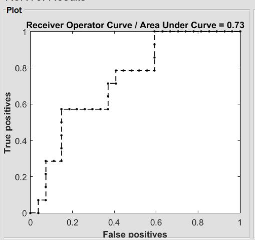
Figure 1 ROC curve of gray matter volume in machine learning to evaluate differences between AIDS group and control group
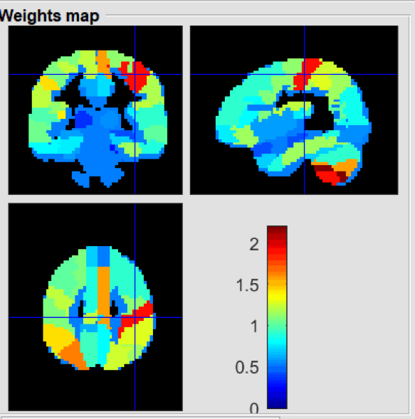
Figure 2 The weight map of gray matter volume obtained from the machine learning demonstrates the volume differences of brain areas between AIDS group and control group. Red color in red means the weighting factor is higher compared to blue color.