1881
Incorporating Deep Learning into Multi-shot EPI DWI Reconstruction1Institute of Science and Technology for Brain-Inspired Intelligence, Fudan University, Shanghai, China, 2Human Phenome Institute, Fudan University, Shanghai, China, 3Philips Healthcare, Shanghai, China, 4Philips Heathcare, Beijing, China
Synopsis
This work tried to optimize MUSE for high resolution multi-shot EPI DWI reconstruction by using Convolutional neural network (CNN). By using multi-scale U-net learning neural network, final reconstructed images showed less artifacts due to the improved phase estimation. Besides, CNN can improve the computational efficiency for the image reconstruction process.
Introduction
Diffusion Weighted Imaging (DWI) has been developed as a powerful technique for clinical neurological disorders disease application1. Multi-shot Echo-Planar Imaging (EPI) based DWI shows great potential for detecting diffusion properties of fine anatomical structures with high spatial resolution and reduced geometry distortion. However, linear and nonlinear phase variation terms often occur among shots due to various motions. Previous studies2,3 have shown that motion-induced artifacts could be reduced if phase inconsistency among different shots can be corrected.Recently, deep learning has been introduced into reconstructions and outperformed conventional MRI reconstruction algorithms. Specifically, recent studies4,5 about k-space deep learning for EPI correction and accelerated MRI using magnitude and phase network showed high potential in image reconstructions.
This work tried to apply machine learning into the framework of multi-shot EPI DWI reconstruction.
Methods
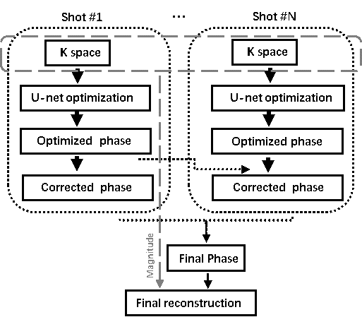
U-net phase correction: By incorporating U-net into MUSE, the whole process of optimized algorithm is shown in Fig. 1. Instead of using SENSE for phase calculation in traditional MUSE, the step of phase estimation was performed by using U-net network with all shots for different diffusion weightings. The calculated phases were used for multi-shot phase correction thereafter.MR Imaging: All MRI experiments were performed on a Philips 3.0 T clinical scanner (Philips Healthcare, Best, the Netherlands). The brain images were acquired from 12 healthy volunteers using a 16-channel phase-array head coil with multi-shot EPI DWI sequence. All DWI data with 4-shot interleaved EPI DWI sequence were acquired from 20 slices, covering whole brain with slice gap of 6 mm. Other parameters were shown as following: FOV=230×230 mm2, acquisition matrix=252×256, special resolution=0.9×0.9×5 mm3, TR/TE=4422/160 ms, scan time ≈400 s, b values =0, 20, 50, 100, 150, 200, 400, 800, 1200 and 2000 s/mm2. Average number for different diffusion weightings were 1, 1, 1, 1, 1, 1, 1, 3, 5, 7, respectively. All subjects were provided by written informed consent.
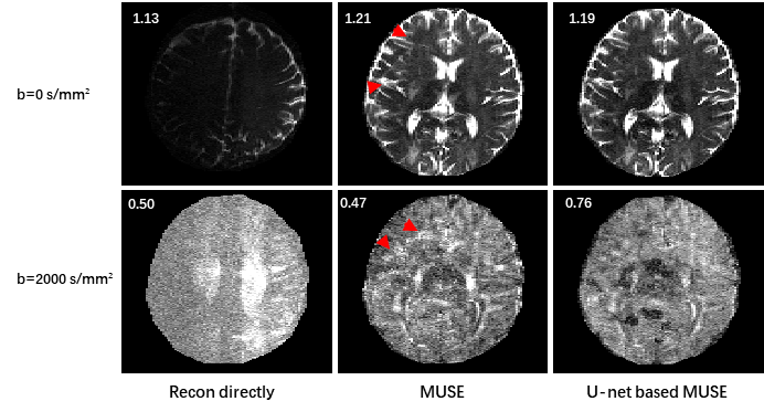
Reconstruction: All images were fully sampled with 4 shots. The performance of U-net based MUSE and traditional method were evaluated. First, phases estimated by traditional MUSE were compared with those by neural network optimized MUSE. Second, final reconstructions of all diffusion weightings from traditional MUSE and the proposed U-net optimization method were shown for comparison.
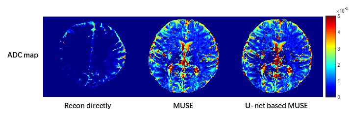
Quantitative analysis:The ADC was obtained by fitting the equation: SIb/SI0=exp(-b×ADC) from the whole brain measurements using a least square nonlinear fitting algorithm in MATLAB (The Mathworks Inc., Natick, MA), where SIb represents signal intensity at different b values.
Results
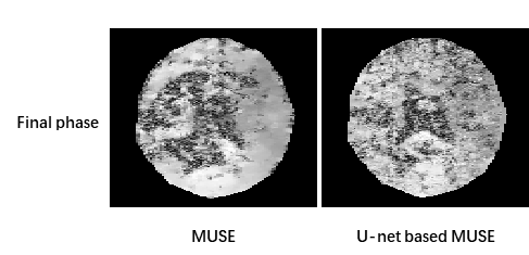
Compared with MUSE reconstruction, U-net based MUSE showed high advantage in alleviating motion induced artifacts, reflected by high SNR performance, which was shown in Fig. 2 (red arrow). Corresponding SNRs were: 1.129 and 0.497, 1.205 and 0.465, 1.186 and 0.757 for b=0 and 2000 s/mm2, respectively.These SNR improvements are reasonable, since they are consistent with physiological structure in central neural regions, especially CSF. Fig.3 illustrated the estimated phase differences between U-net based MUSE and traditional MUSE method.
In quantitative comparison, the ADC of U-net training method showed comparative results when compared with traditional MUSE. U-net shows advantage in some regions when severe field inhomogeneity occurred (Fig. 4).
Discussion
Although U-net based method showed improvements compared to traditional MUSE, under some circumstances, for instance region with high field inhomogeneity or high b-values with serious signal loss, the results haven’t been improved or even more blurred.Conclusion
The proposed U-net based multi-shot EPI DWI methods can improve the performance of MUSE by optimizing the phase correction process.Acknowledgements
This work was supported by Shanghai Municipal Science and Technology Major Project (No.2017SHZDZX01), Shanghai Municipal Science and Technology Major Project (No.2018SHZDZX01) and ZJLab, Shanghai Natural Science Foundation (No. 17ZR1401600) and the National Natural Science Foundation of China (No. 81971583).References
1. Moseley ME, Cohen Y, Kucharczyk J, et al. Diffusion-weighted MR imaging of anisotropic water diffusion in cat central-nervous-system. Radiology. 1990;176;439-45.
2. Nan-kuei Chen, et al. A robust multi-shot scan strategy for high-resolution diffusion weighted MRI enabled by multiplexed sensitivity-encoding (MUSE). Magn Reson Med.2013;72;41-7.
3. Zhe Zhang, et al. Self-feeding MUSE: A Robust Method for High Resolution Diffusion Imaging Using Interleaved EPI. NeuroImage. 2015;
4. Dongwook Lee, et al. Deep Residual Learning for Accelerated MRI Using Magnitude and Phase Networks. IEEE transactions on imaging processing. 2018; 65;1985-198.
5. Yoseob Han, et al. Deep learning with domain adaptation for accelerated projection-reconstruction MR. Magn Reson Med.2018;80;1189-205.
Figures