1849
Brain GABA levels distinguish multiple sclerosis and neuromyelitis optica using edited magnetic resonance spectroscopy1Shandong Medical Imaging Research Institute, Shandong University, Jinan, China, 2Philips Healthcare, shanghai, China
Synopsis
Distinguishing relapsing-remitting multiple sclerosis (RRMS) and neuromyelitis optica (NMO) is clinically important because they differ in prognosis and treatment. Edited magnetic resonance spectroscopy (MRS), using the MEGA-PRESS sequence, is the most widely used technique for detecting GABA in the human brain. However, none of the existing studies are performed to compare the brain GABA levels between patients with MS and NMO. In this study, we found GABA levels differences in the LHC and PCC between the RRMS and NMO patients, which may be helpful in distinguishing RRMS from NMO after further validation.
Introduction
Although immunological, clinical, and imaging measures have exhibited the potential to distinguish relapsing-remitting multiple sclerosis (RRMS) and neuromyelitis optica (NMO), no method has been able to completely differentiate MS from NMO. Thus, there is an urgent need to identify imaging markers that could be used to distinguish these two disorders. Gamma-aminobutyric acid (GABA) is the main inhibitory neurotransmitter in the central nervous system. Previous studies have demonstrated that there is a dysfunctional GABAergic neurotransmission in animal models of multiple sclerosis.[1] Edited magnetic resonance spectroscopy (MRS), using the MEGA-PRESS sequence, is the most widely used technique for detecting GABA in the human brain. However, none of the existing studies are performed to compare the brain GABA levels between patients with MS and NMO.Material and Methods
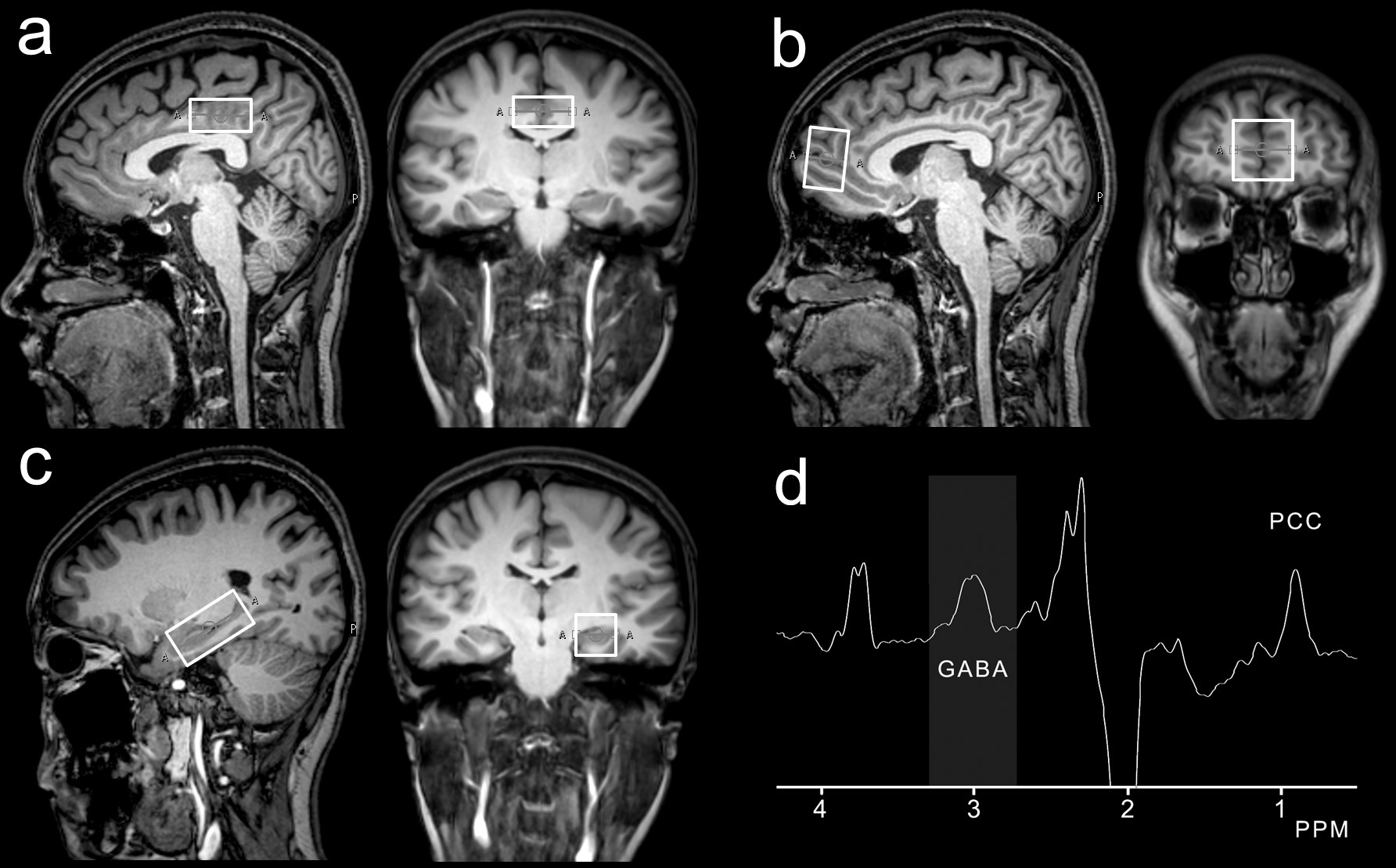
39 patients with RRMS (27 males/12 females, mean age 43.7±4.9 years) and 35 patients with NMO (25 males/10 females, mean age 43.1±3.9 years) and 40 healthy controls (28 males/12 females, mean age 43.8±4.1 years) were examined on a 3T scanner and T1-weighted three-dimensional TFE images were used as a localizer. The MEGA-PRESS sequence (TR 2000 ms; TE 68 ms; 256 averages) was used to measure GABA concentrations in the posterior cingulate cortex (PCC), medial prefrontal cortex (mPFC) and left hippocampus (LHC). For quantification, a shorter measurement (8 averages) of the unsuppressed water signal was obtained. The MRS data were analyzed using 'Gannet' (GABA-MRS Analysis Tool) in Matlab with Gaussian curve fitting to the GABA peaks.[2] 3 Hz exponential line broadening was applied. The ratios of the integrals of the GABA and water signals, making corrections for T1 and T2 relaxation times and partial volume effects, were used to calculate water-scaled GABA concentration in mmol/L (mM) using a formula.Results
The RRMS group showed reduced GABA levels in the PCC and LHC compared to the control group (LHC: 1.20 ± 0.23 mM vs. 1.36 ± 0.23 mM, p = 0.026; PCC: 1.13 ± 0.13 mM vs. 1.22 ± 0.14 mM, p = 0.02). Importantly, The RRMS group showed reduced GABA levels in the PCC and LHC compared to the NMO group (LHC: 1.20 ± 0.23 mM vs. 1.40 ± 0.36 mM, p = 0.035; PCC: 1.13 ± 0.13 mM vs. 1.28 ± 0.16 mM, p = 0.003). No statistical difference in GABA concentrations in the MPFC region was seen among three groups.Conclusion
Distinguishing relapsing-remitting multiple sclerosis (RRMS) and neuromyelitis optica (NMO) is clinically important because they differ in prognosis and treatment. In this study, we found GABA levels differences in the LHC and PCC between the RRMS and NMO patients, which may be helpful in distinguishing RRMS from NMO after further validationAcknowledgements
This work was supported by the National Natural Science Foundationof China for Young Scholars (no. 81601479) and Tai Shan Scholars Project (No.tsqn201812147).References
1. Gilani, A. A., Dash, R. P., Jivrajani, M. N., Thakur, S. K., & Nivsarkar, M.(2014). Evaluation of GABAergic transmission modulation as a novelfunctional target for Management of Multiple Sclerosis: Exploringinhibitory effect of GABA on glutamate-mediated excitotoxicity.Advances in Pharmacological Sciences, 2014, 632376.
2. Edden, R. A., Puts, N. A., Harris, A. D., Barker, P. B., & Evans, C. J. (2014).Gannet: A batch-processing tool for the quantitative analysis ofgamma-aminobutyric acid-edited MR spectroscopy spectra. Journal ofMagnetic Resonance Imaging, 40(6), 1445–1452.
Figures