1834
Comparison between MPRAGE GRAPPA 2×1 with MPRAGE Wave-CAIPI 3×31Department of Diagnostic Imaging and Nuclear Medicine, Kyoto University Graduate School of Medicine, Kyoto, Japan, 2Human Brain Research Center, Kyoto University Graduate School of Medicine, Kyoto, Japan, 3Siemens Healthineers AG, Shenzhen, China
Synopsis
This comparison study between MPRAGE with GRAPPA and MPRAGE with Wave-CAIPI has been performed at the clinical MR scanner. MPRAGE with Wave-CAIPI 3×3 shows relatively good contrast despite its short scan time of 1 m 42 s. No segmentation error was found in MPRAGE with Wave-CAIPI 3×3 in our cohort. The correlation of cerebral cortex and cerebral white matter was very high between 2 MPRAGEs. In deep gray matters except pallidum and hippocampus, the correlation of VOIs were also high. These results suggest that volumes derived from automated segmentation of MPRAGE with Wave-CAIPI are reliable measures.
Introduction
By taking advantage of coil sensitivity encoding from multi-channel receiver coils, reduction of phase-encoding steps has been available with the parallel imaging technique such as SENSE and GRAPPA. Controlled aliasing in parallel imaging results in higher acceleration (2D-CAIPIRINHA) has enabled image reconstruction with further acceleration, which can efficiently employ the sensitivity variation in phase encoding directions [1]. To further utilize the sensitivity information in the readout direction, bunch phase encoding (BPE) has been combined with parallel imaging to improve the image reconstruction [2-3]. Wave-CAIPI acquisition extends and combines the BPE and 2D-CAIPIRINHA by playing sinusoidal gradients during the readout of each phase encoding line and introducing interslice shifts across the aliasing slices [4-5], which enables highly accelerated volumetric imaging with low artifact and signal-to-noise ratio (SNR) penalties. More acceleration has been expected with the introduction of Wave-CAIPI, however, image quality has not been evaluated well in the clinical practices.The purpose of this study is to evaluate MPRAGE with 9-fold acceleration by using Wave-CAIPI (Wave-CAIPI 3×3) in clinical scanners, and to compare MPRAGE Wave-CAIPI 3×3 with conventional MPRAGE GRAPPA 2×1.
Method
- Subjects
- MR imaging
- Post-imaging process
Results
No segmentation error was found in both MPRAGEs in our cohort. Representative images of original MPRAGE and segmented images by using recon-all function of FreeSurfer were shown in Figure 1 and 2. MPRAGE GRAPPA 2×1 showed better image quality, however, MPRAGE Wave-CAIPI 3×3 shows relatively good contrast between gray and white matters in healthy subjects (Figure 1). Of note, MPRAGE Wave-CAIPI 3×3 also showed nice segmented results even in patients with brain atrophy (Figure 2).We compared the volumes of each VOIs between GRAPPA 2×1 and Wave-CAIPI 3×3. The correlation of all VOIs was 0.991 [0.989 - 0.992] (Figure 3). Volumes of each VOI and the correlation between GRAPPA 2×1 and Wave-CAIPI 3×3 are shown in Figure 4 and 5. The results of cerebral white matter and cerebral cortex were shown in Figure 4. The correlation of cerebral cortex and cerebral white matter was very high as 0.98-0.99. The results of deep gray matters, hippocampus and amygdala were shown in Figure 5. The correlation of almost VOIs were higher than 0.9.
Discussion
Wave-CAIPI MPRAGE was evaluated in comparison with other accelerated techniques of MPRAGE. MPRAGE with Wave-CAIPI 3×3 shows relatively better contrast despite of its short scan time. Less imaging noise and segmentation error was found in MPRAGE with Wave-CAIPI 3×3.Wave-CAIPI showed good image quality for its high acceleration factor. Short scan time of Wave-CAIPI can reduce motion artifact and high SNR derived from 64-channel coil may attribute to relatively good image quality.
The correlation of cerebral cortex and cerebral white matter was very high as 0.98-0.99. From our result, we can completely substitute MPRAGE GRAPPA 2×1 with MPRAGE Wave-CAIPI 3×3 for volumetric evaluation of cerebral white matter and cerebral cortex.
In deep gray matters except pallidum and hippocampus, the correlation of VOIs were higher than 0.9. The correlation of pallidum and amygdala were relatively lower than that of other VOIs. This is probably because pallidum and amygdala are relatively small in volume. In addition, pallidum and amygdala show less image contrast on both MPRAGE GRAPPA 2×1 and Wave-CAIPI 3×3, therefore, parcellation of these VOIs were derived from probabilistic image segmentation.
The structural volumes of MPRAGE Wave-CAIPI 3×3 were almost similar to MPRAGE GRAPPA 2×1 in average. SNR should be lower in Wave-CAIPI 3×3, compared with Grappa 2×1. However, based on our results of FreeSurfer, putative lower SNR of Wave-CAIPI seems to have no impact on segmentation.
Conclusion
This comparison study between MPRAGE with GRAPPA and MPRAGE with Wave-CAIPI has been performed at the clinical MR scanner. MPRAGE with Wave-CAIPI 3×3 shows relatively better contrast despite of its short scan time of 1 m 42 s. No segmentation error was found in MPRAGE with Wave-CAIPI 3×3. The correlation of cerebral cortex and cerebral white matter was very high. In deep gray matters except pallidum and hippocampus, the correlation of VOIs were high. These results suggest that volumes derived from automated segmentation of MPRAGE with Wave-CAIPI are reliable measures.Acknowledgements
We are grateful to Mr. Yuta Urushibata, Siemens Healthcare K. K., for helpful discussions.References
- Breuer F, Blaimer M, Mueller MF, Seiberlich N, Heidemann RM, Griswold MA, Jakob PM. Controlled aliasing in volumetric parallel imaging (2D CAIPIRINHA). Magn Reson Med 2006;55.3:549–556.
- Moriguchi H, Duerk JL. Magn Reson Med. 2006 Mar;55(3):633-48. Bunched phase encoding (BPE): a new fast data acquisition method in MRI.
- Breuer FA, Moriguchi H, Seiberlich N, Blaimer M, Jakob PM, Duerk JL, Griswold MA. Magn Reson Med. 2008 Aug;60(2):474-8. Zigzag sampling for improved parallel imaging.
- Bilgic B, Gagoski BA, Cauley SF, Fan AP, Polimeni JR, Grant PE, Wald LL, Setsompop K. Magn Reson Med. 2015 Jun;73(6):2152-62. Wave-Caipi for highly accelerated 3D imaging.
- Daniel P., Kawin S., Stephen F. Magn Reson Med. 2018 Jan; 79(1): 401–406. Wave-CAIPI for Highly Accelerated MP-RAGE Imaging.
- Dale, A.M., Fischl, Bruce, Sereno, M.I. Neuroimage. 1999 Feb;9(2):179-94. Cortical Surface-Based Analysis I: Segmentation and Surface Reconstruction.
Figures
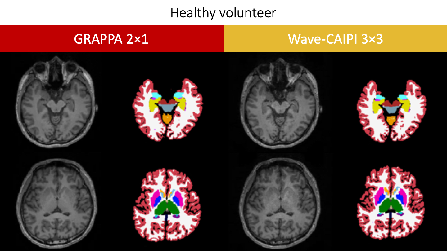
Fig. 1. A healthy subject.
Representative images of original MPRAGE and segmented images by using recon-all function of FreeSurfer were shown in Figure 1.
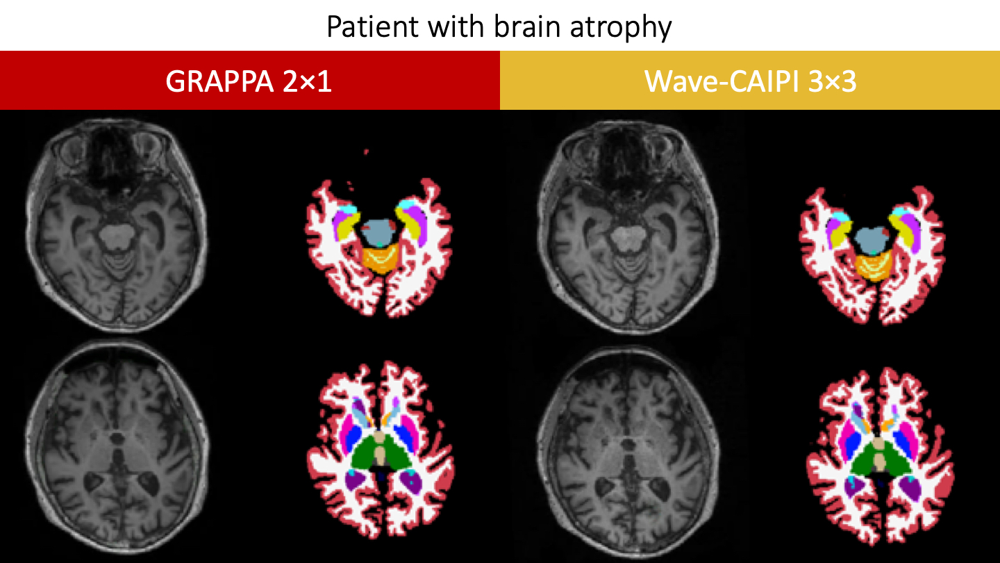
Fig. 2. A patient with brain atrophy.
Representative images of original MPRAGE and segmented images by using recon-all function of FreeSurfer were shown in Figure 2.
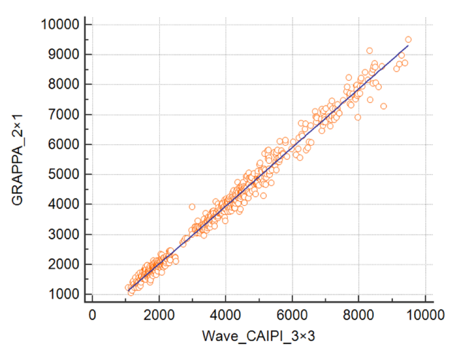
Fig. 3
The correlation of all VOIs between GRAPPA 2×1 and Wave-Caipi 3×3 was 0.991 [0.989 - 0.992].
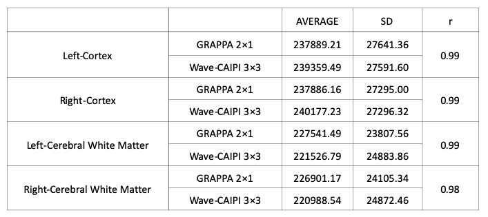
Fig. 4
Volumes of each VOI and the correlation between GRAPPA 2×1 and Wave-Caipi 3×3 are shown in Figure 4. The results of cerebral white matter and cerebral cortex were shown in Figure 4.
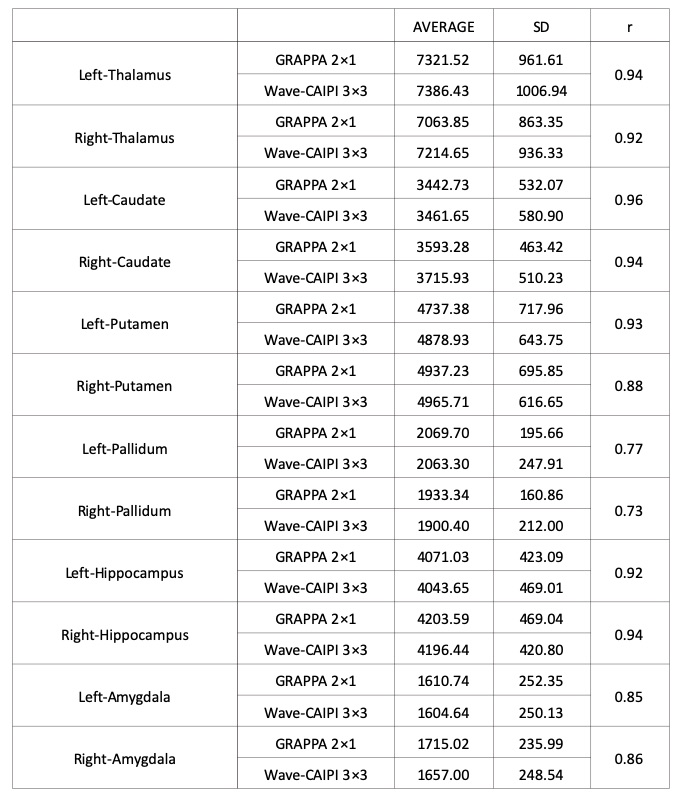
Fig. 5
Volumes of each VOI and the correlation between GRAPPA 2×1 and Wave-Caipi 3×3 are shown in Figure 5. The results of deep gray matters, hippocampus and amygdala were shown in Figure 5.