1821
Pointwise Encoding Time Reduction with Radial Acquisition in Subtraction-Based MRA: Assessing Intracranial Saccular Aneurysms
Qing Fu1, Xiao-yong Zhang2, Ding-xi Liu1, and Xian-bo Deng1
1Department of Radiology, Union Hospital, Tongji Medical College, Huazhong University of Science and Technology, Wuhan, China, 2MR Collaborations, Siemens Healthcare Ltd., Shenzhen, China
1Department of Radiology, Union Hospital, Tongji Medical College, Huazhong University of Science and Technology, Wuhan, China, 2MR Collaborations, Siemens Healthcare Ltd., Shenzhen, China
Synopsis
This study compared the clinical utility of the pointwise encoding time reduction with radial acquisition (PETRA) sequence in subtraction-based magnetic resonance angiography (MRA) with conventional time-of-flight MRA (TOF-MRA) for evaluating intracranial saccular aneurysms. The results showed no substantial differences between PETRA-MRA and TOF-MRA in detecting, localizing, and determining the size of aneurysms. Furthermore, PETRA-MRA produced better image quality and provided a higher correlation with computed tomography angiography (CTA) in assessing morphologic parameters of aneurysms than TOF-MRA.
Introduction
While time-of-flight magnetic resonance angiography (TOF-MRA) is widely used for imaging intracranial saccular aneurysms, it is limited by saturation effects and dephasing artifacts, which may lead to substantial loss of signal within the vessels. The ultrashort echo-time pointwise encoding time reduction with radial acquisition (PETRA) technique has the potential to be resistant to flow dephasing. The aim of this study was to investigate the clinical utility of PETRA with subtraction-based MRA (PETRA-MRA) for assessing intracranial saccular aneurysms compared with TOF-MRA on a 3T MR scanner.Methods
The IRB-approved MR examinations were performed on a 3T MR scanner (Magnetom Skyra, Siemens Healthcare, Germany), equipped with 45mT/m achievable gradient strength and 200T/m/s maximum slew rate, using a 20-channel head-neck coil. Forty patients (12 male and 28 female; mean age, 52 years; age range, 33-70 years) with intracranial saccular aneurysms identified on 3D-TOF-MRA were enrolled in the study.PETRA-MRA was performed on each patient without contrast material within< 24 hours. Eleven of the enrolled patients (1 male and 10 female; mean age, 53 years; age range, 43-62 years) underwent intracranial computed tomography angiography (CTA). 3D-TOF-MRA was performed in an axial orientation with the following parameters: field-of-view (FOV)=200x200mm2, repetition time/echo time (TR/TE)=20.00/3.43ms, flip angle=18°, imaging matrix = 320x256, acquired voxel size=0.79x0.63x1.00mm3, bandwidth = 186Hz/Px,acquisition time(TA)=3min, 45sec. For PETRA-MRA in the same section orientation, the following imaging parameters were used: FOV=256x256 mm2,TR/TE=3.54/0.07ms, flip angle=6 °,imaging matrix = 352x352, acquired voxel size=0.73x0.73x0.73mm3, bandwidth=369Hz/Px, radial views number=75000. Since PETRA-MRA subtracts from two imaging datasets with and without a slice-selective saturation slab in the bottom of the imaging volume, the acquisition time for PETRA-MRA was 4min,36sec for data without a saturation band, and 7min,45sec for data with a saturation band placed at the bottom of the imaging volume.The morphologic parameters, including the maximum diameter of the aneurysm, mean diameter of the aneurysm neck, mean diameter of the parent artery, inflow-angle between the aneurysm and parent artery, aspect ratio (AR, dome-to-neck ratio) and size ratio (SR, dome-to-parent artery diameter ratio) were measured and calculated using PETRA-MRA, TOF-MRA, and CTA. Two radiologists independently scored the quality of PETRA-MRA and TOF-MRA images. Signal homogeneity and sharpness of images were scored using a 4-point scale: 4, excellent (excellent-quality diagnostic information with homogeneous signals within aneurysms); 3, good (good-quality diagnostic information with minimal blurring or artifacts within aneurysms); 2, poor (structures were slightly visible but with significant blurring or artifacts within aneurysms, not diagnostic); and 1, not assessable (impossible to make diagnosis).The acoustic noise levels of PETRA-MRA and TOF-MRA were recorded using a decibel meter placed laterally 2 meters from the MR scanner.The image quality and acoustic noise levels of PETRA-MRA and TOF-MRA were compared using the Wilcoxon signed rank test. Intraclass correlation coefficient (ICC) estimates and 95% confident intervals (CIs) were calculated based on absolute agreement, using a two-way random model for the inter-observer reliability analysis (< 0.50, poor reliability; 0.5 to 0.75, moderate reliability; 0.75 to 0.9,good reliability; >0.9, excellent reliability). P < 0.05 was considered significant. Pearson correlation coefficient (r) was used to evaluate the correlations of PETRA-MRA and TOF-MRA with CTA.
Results
The detection and localization of all 44 aneurysms identified by PETRA-MRA were 100% consistent with TOF-MRA. The most common sites of aneurysms were at the C6 segment (20/44), followed by the C7 segment (7/44).There was no significant difference detected in aneurysm sizes between PETRA-MRA and TOF-MRA(P<0.01).The morphologic parameters evaluated on PETRA-MRA were more closely correlated with CTA than those assessed on TOF-MRA(Table 1).The subjective image score of PETRA-MRA (3.86±0.35) was significantly higher than that of TOF-MRA (3.51±0.55) (z=-3.500, P<0.001) (Figs. 1 and 2).Inter-observer reliability was excellent for both PETRA-MRA (ICC 0.90, 95% CI 0.82-0.94, P <0.001) and TOF-MRA (ICC 0.96, 95% CI0.93-0.98, P <0.001). The acoustic noise level of PETRA-MRA was significantly lower than that of TOF-MRA (59 dB verus 73 dB, P<0.01).
Discussion and Conclusion
PETRA-MRA is an ultrashortecho time (UTE) technique (TE=0.07ms), that combines half-projection radial acquisition in outer k-space and single pointwise measurement on a Cartesian trajectory in center k-space, which renders it immune to flow dephasing artifacts1,2.Our findings support the utility of PETRA-MRA for detecting and localizing intracranial saccular aneurysms. Additionally, compared with TOF-MRA, PETRA-MRA provides superior image quality, higher correlation with CTA for assessing morphologic parameters, and a lower acoustic noise level.Acknowledgements
noneReferences
1. Grodzki DM, Jakob PM, Heismann B. Ultrashort echo time imaging using pointwise encoding time reduction with radial acquisition (PETRA). Magn Reson Med 2012;67:510-518
2. Natsuaki Y, Bi X, Grodzki DM, et al. PETRA qMRA: Towards Zero-Flow dephasing intracranial Non-Contrast MR angiography. In:Proc. Intl. Soc. Mag. Reson. Med., 2015.
Figures
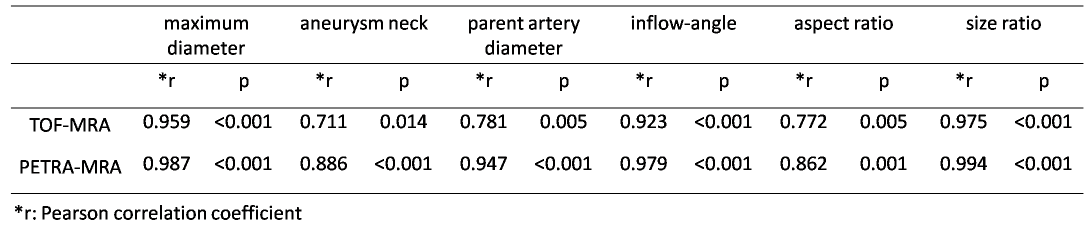
Table 1. Correlations of PETRA-MRA and TOF-MRA with CTA for evaluation of morphologic parameters of intracranial saccular aneurysms.

Figure 1. The location and morphologic parameters of a small aneurysm were identified clearly by both TOF-MRA and PETRA-MRA.

Figure 2. The signal homogeneity within the aneurysm and correlation of morphological parameters with CTA were better for PETRA-MRA than for TOF-MRA.