1819
Microscopic fractional anisotropy measurements from double diffusion encoding for human brain tumor diagnosis1Toho University Omori Medical Center, Tokyo, Japan, 2Radiology, Juntendo University School of Medicine, Tokyo, Japan, 3Research and Development Center, Canon Medical Systems Corporation, Kanagawa, Japan, 4Advanced MRI development PJ Team, Canon Medical Systems Corporation, Kanagawa, Japan, 5Juntendo University Hospital, Tokyo, Japan, 6Canon Medical Systems Corporation, Tokyo, Japan, 7Radiology, The University of Tokyo Graduate School of Medicine, Tokyo, Japan
Synopsis
We investigated the values and image contrast of μFA using double diffusion encoding in patients with brain tumors in vivo, as a preliminary study. In general, μFA showed higher values than FA did in the brain tumors. There is no correlation among μFA and FA and ADC, in the meningiomas and solid part of glioblastoma and malignant lymphoma. However, more investigation will be needed to establish the usefulness of μFA in clinical use.
Introduction
Recently, microscopic fractional anisotropy (μFA) maps generated from double diffusion encoding (DDE) technique1 showed a promising results to investigate the microstructural changes in the pathologic lesions (such as demyelinated lesion and brain tumors) in vivo2, 3. However, little has been reported the values and image contrast of μFA maps in several kinds of brain tumors. The purpose of this study is to investigate and clarify the values and image contrast of μFA in patients with brain tumors in vivo, as a preliminary study.Methods
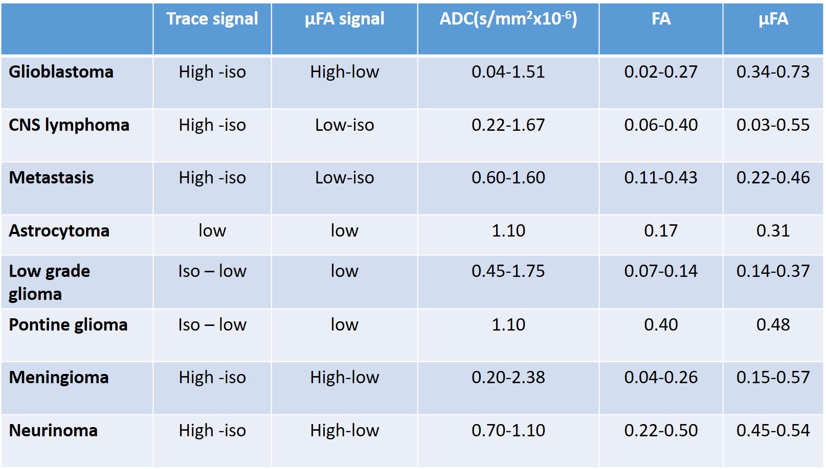
In this prospective study, we enrolled 19 brain neoplasm patients (5 meningioma, 3 acoustic neurinoma, 3 glioblastoma, 2 low grade glioma, 2 metastatic brain tumor, 2 malignant lymphoma, 1 astrocytoma and 1 pontine glioma). After conventional MR imaging including T2-, T1- and occasionally contract enhanced T1-weighted imaging, DDE MR imaging data were acquired on a Vantage Galan 3T/ZGO (Canon Medical Systems Corp.) Imaging parameters for DDE were as follows: repetition time (TR)/echo time, 6600/116 (ms/ms); number of signals acquired, one; section thickness, 3 mm; 42 slices; in-plane pixel size, 3.0x 3.0 mm; multiband factor, 2; δ=19, Δ=21, mixing time=32 ms; imaging time, approximately 6 min; 2 b values (1000 and 1000 s/mm 2) with one b=0 image and diffusion encoding in 48 directions, reduced form 72 direction originally reported1. All diffusion MRI data was transferred to an offline workstation and processed using in-house developed software in Matlab (R2017a, Math Works, Inc, Natick, MA) to derive and measure parametric maps of apparent diffusion coefficient (ADC), fractional anisotropy (FA), and µFA. Moreover, the correlation among diffusion metrics were investigated in brain tumors.Results
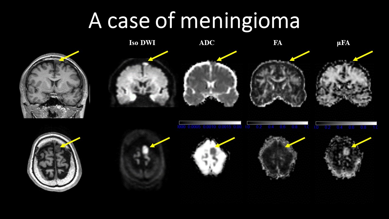
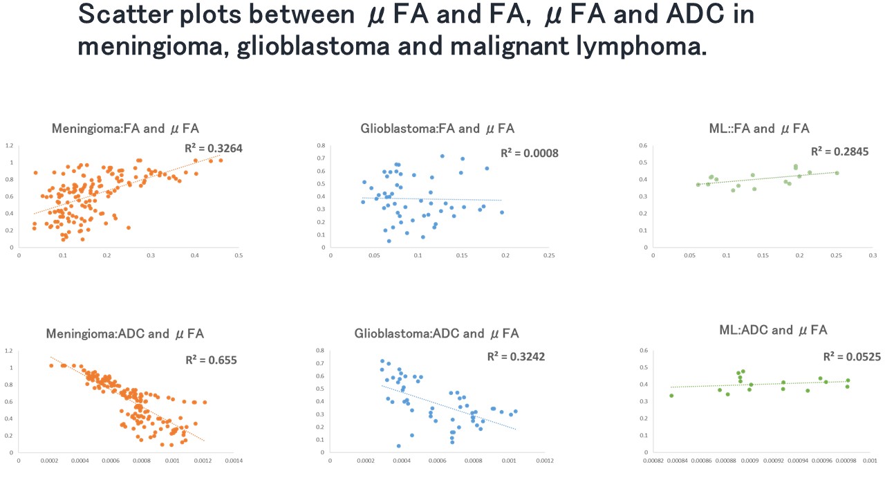
All metrics values and image contrasts were summarized in table 1. In general, μFA showed higher values than FA did in the brain tumors (Figure 1). There is no correlation among μFA and FA and ADC, in the meningiomas and solid part of glioblastoma and malignant lymphoma (table2).Discussion
Double diffusion encoding provides novel contrast based on microscopic diffusion anisotropy of each element in a voxel. The μFA measurements can provide an additional information in vivo brain tumors, compared with conventional diffusion metrics of FA and ADC. For example high μFA and low FA in the tumor indicate the spindle type tumor cell, in case of meningiomas and neurinomas, compatible with histologic knowledge. However, more investigation will be needed to establish the usefulness of μFA in clinical use.Acknowledgements
This work was supported by JSPS KAKENHI Grant Number 19K08161.References
1. Jespersen, Sune Nørhøj, et al. "Orientationally invariant metrics of apparent compartment eccentricity from double pulsed field gradient diffusion experiments." NMR in Biomedicine 26.12 (2013): 1647-1662.
2. Yang, Grant, et al. "Double diffusion encoding MRI for the clinic." Magnetic resonance in medicine 80.2 (2018): 507-520.
3. Szczepankiewicz F, et al. Quantification of microscopic diffusion anisotropy disentangles effects of orientation dispersion from microstructure: applications in healthy volunteers and in brain tumors. Neuroimage. 2015 1;104:241-52.
Figures