1813
Evaluation of Ultrafast Wave-CAIPI 3D FLAIR Versus Standard 3D FLAIR for Quantitative Analysis of White Matter Lesions1Athinoula A. Martinos Center for Biomedical Imaging, Department of Radiology, Massachusetts General Hospital, Boston, MA, United States, 2Faculty of Medicine, Siriraj Hospital Mahidol Univerity, Bangkok, Thailand, 3Department of Radiology, Massachusetts General Hospital, Boston, Massachusetts, Boston, MA, United States, 4Harvard Medical School, Boston, MA, United States, 5Harvard-MIT Division of Health Sciences and Technology, Massachusetts Institute of Technology, Cambridge, MA, United States, 6Department of Physics and Astronomy, Heidelberg University, Heidelberg, Heidelberg, Germany, 7Siemens Healthineers AG, Erlangen, Erlangen, Germany, 8Siemens Shenzhen Magnetic Resonance Ltd., Shenzhen, Shenzhen, China
Synopsis
Quantification of cerebral white matter lesion volume has become increasingly feasible for routine clinical evaluation and research due to the availability of automated segmentation tools and 3D FLAIR sequences. However, these sequences suffer from long acquisition times, limiting their widespread use. We demonstrate that quantitative white matter lesion volumes estimated using ultrafast Wave-CAIPI SPACE-FLAIR obtained in <3 minutes show excellent agreement with standard SPACE-FLAIR requiring >7 minutes of scanning in patients undergoing clinical evaluation for suspected MS and epilepsy. Wave-CAIPI SPACE-FLAIR may facilitate the adoption of 3D FLAIR sequences for lesion evaluation in patients with MS and other white matter diseases.
Introduction
Quantification of cerebral white matter lesion volume has become increasingly feasible for routine clinical evaluation and use in clinical trials due to the availability of automated segmentation tools and three-dimensional fast spin echo fluid-attenuated inversion recovery (3D FSE FLAIR) sequences, which delineate cerebral white matter lesions at high isotropic resolution. However, these sequences suffer from long acquisition times, which limit their widespread use. The goal of this study was to evaluate an ultrafast Sampling Perfection with Application optimized Contrasts by using different flip angle Evolutions (SPACE) FLAIR sequence using Wave-CAIPI encoding (Wave SPACE-FLAIR) [1,2] compared to standard SPACE-FLAIR for quantitative analysis of cerebral white matter lesions in a clinical setting.Methods
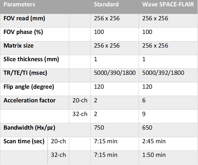
Data acquisition:This study was approved by the IRB and was HIPAA compliant. Forty-six consecutive patients undergoing brain MRI as part of routine clinical work-up and/or surveillance for multiple sclerosis and epilepsy were enrolled. MRI scans were performed on one of two clinical 3T MR scanners (MAGNETOM Prisma, Siemens Healthcare, Erlangen, Germany) using 20- or 32-channel multi-array receiver coils, depending on the fit and comfort of the patient. Each scan included a standard 3D SPACE-FLAIR (acceleration factor R=2, scan time TA=7:15 min) and resolution-matched ultrafast 3D Wave SPACE-FLAIR (R=6, TA=2.45 min for the 20-ch coil and R=9, TA=1:50 min for the 32-ch coil) sequences. Detailed acquisition parameters for the standard and Wave SPACE-FLAIR sequences are shown in Table 1.
Images were reviewed in a blinded fashion for motion and presence of lesions by an experienced neuroradiologist (C.N., 9 years of experience). Patients were excluded if either set of images showed moderate to severe motion or if no white matter lesions were detected.
White matter lesion analysis:
Cerebral white matter lesions were segmented using the lesion prediction algorithm (LPA) implemented in the Lesion Segmentation Tool (LST) toolbox version 2.0.15 (www.statistical-modelling.de/lst.html) in the SPM. Lesion probability maps generated by LPA from the standard and Wave SPACE-FLAIR sequences were compared using the longitudinal pipeline in LST. Binarized lesion maps were created based on the lesion probability maps derived from standard and Wave SPACE-FLAIR sequences using threshold values adjusted by the radiologist to exclude voxels outside the brain parenchyma and within normal gray matter.
Pearson’s correlation coefficients, absolute symmetrized percent change (ASPC), and Dice similarity coefficients were used to compare quantitative volumetric measurements between sequences.
Results
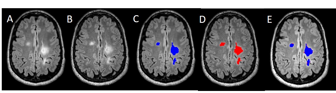
Of the 46 patients scanned, 23 patients with white matter lesions (18 (78.3%) female, mean age 49 years (range 19-86), 11 MS and 12 epilepsy) were included in the quantitative evaluation. 56% of patients were scanned with the 20-channel coil. Standard and Wave-CAIPI SPACE-FLAIR sequences showed excellent correlation of lesion volumes segmented by LST (r=0.99, p<0.0001). The mean Dice similarity coefficient for white matter lesions was 0.99±0.02 (range 0.91 to 1). The mean ASPC was -0.04 ±0.21% (range -0.98 to 0.14%) between the two sequences. (Figure 1).Discussion and Conclusion
Quantitative white matter lesion volumes estimated using ultrafast Wave SPACE-FLAIR and LST obtained in less than three minutes showed excellent agreement with standard SPACE-FLAIR requiring over seven minutes of scan time in patients undergoing clinical evaluation for MS and epilepsy. These findings may facilitate the increased use of 3D FLAIR sequences in patients with MS and other white matter diseases.Acknowledgements
This work was supported by the National Institute of Biomedical Imaging and Bioengineering of the National Institutes of Health under award number R01EB020613 and by a research grant from Siemens Healthineers.References
1. Polak D, Setsompop K, Cauley SF, et al. Wave-CAIPI for highly accelerated MP-RAGE imaging. Magnetic resonance in medicine 2018;79:401-406.
2. Polak D, Cauley S, Huang SY, et al. Highly-accelerated volumetric brain examination using optimized wave-CAIPI encoding. Journal of magnetic resonance imaging : JMRI 2019;50:961-974.
Figures