1811
Optimized Structural Imaging at 7T using ME-MP2RAGE1German Center for Neurodegenerative Diseases, Bonn, Germany, 2Center for Neuroscience Imaging Research, Institute for Basic Science & Department of Biomedical Engineering, Sungkyunkwan University, Suwon, Korea, Republic of, 3Techna Institute & Koerner Scientist in MR Imaging, University Health Network, Toronto, ON, Canada, 4Department of Physics and Astronomy, University of Bonn, Bonn, Germany
Synopsis
In this work, we present an optimized, scan-time efficient multi-echo MP2RAGE sequence (ME-MP2RAGE) for improved brain segmentation at ultra-high field. Apart from removing B1-, M0, and T2* dependencies (MP2RAGE features), the proposed method removes susceptibility induced image distortions along the readout direction. Additionally, an optimized reordering scheme was implemented for scan-time optimization.
Introduction
In the last years, UHF (7T) imaging has become an increasingly important modality for neuroscience applications. However, increased B0 and B1 inhomogeneities reduce image quality and induce artifacts. To overcome these issues in T1-weighted structural imaging, the MP2RAGE sequence was introduced, removing the reception field bias and partially the transmit field inhomogeneity [1]. Image distortions arising from susceptibility differences are not accounted for. These distortions induce systematic errors during analysis of cortical thickness and brain volume estimation in quantitative brain morphometry. To overcome susceptibility induced image distortions a multi-echo MP2RAGE sequence was implemented, employing high bandwidth signal reception per contrast and echo [2]. Furthermore, the sequence sampling pattern was optimized for imaging efficiency.Methods
All experiments were performed on a Terra 7T scanner (Siemens Healthineers, Erlangen, Germany) using a head array coil with 32 receive channels (Nova Medical, Wilmington, USA). Images were acquired using an optimized ME-MP2RAGE sequence (two inversion contrasts, four echoes each inversion contrast). To minimize distortions in readout direction a high sampling bandwidth was chosen. Standard MP2RAGE image reconstruction was performed on the individual echoes for both contrasts. For the final image, all four echoes were averaged to obtain an image with SNR compared to a standard low bandwidth acquisition.To keep scan time as short as possible, while simultaneously maintaining image contrast, an optimized linear reordering scheme with elliptical scanning was used [3]. It allows to choose the turbofactor (number of excitation pulses per TR) independent of the phase and partition encoding.
Imaging was performed in one healthy volunteer. ME-MP2RAGE sequence parameters: 4 echoes (bipolar readout), TR = 4730 ms, TI1/TI2 = 840 / 2370 ms, BW= 1120 Hz/px, turbofactor 156, 0.8 mm resolution isotropic, matrix size 320x320x192 (whole brain coverage), CAIPIRINHA (R=3, Δ=1), total scan time ≈ 8 min.To demonstrate the effect of the receiver bandwidth on image distortions, two MP2RAGE scans were acquired with a standard "low" readout bandwidth (250 Hz/px, second scan acquired with reversed readout direction). Both MP2RAGE scans were co-registered to each other using a rigid body mid-way transformation [4]. Afterwards a difference image was calculated.The insensitivity of ME-MP2RAGE against off-resonances in readout direction is shown by calculating two different sets of composite images, combining odd and even echoes as follows:
Composite image 1: (1st + 3rd) - (2nd + 4th) echo.
Composite image 2: (1st + 2nd) - (3rd + 4th) echo.
Results
Figure 1 shows the MP2RAGE difference images (left), subtracting the acquisition two (reversed readout direction) from acquisition one. To better visualize the locally induced image distortions, a corresponding animated GIF of the individual images is depicted as well. Especially in regions with large off-resonances, severe distortions are visible.In contrast, the ME-MP2RAGE difference images (see figure 2) suffer less from susceptibility induced image distortions in readout direction. Combining odd and even echoes in two separate images yields the optimal result in terms of distortion reduction (figure 2, right).Discussion
It is shown that ME-MP2RAGE enhances image quality in 3D whole brain T1 weighted imaging at 7T. Image distortions in readout direction induced by static field inhomogeneities are minimized. Hence, a more precise brain segmentation and cortical thickness estimation is possible. As total data sampling time during readout is equal, only a slight SNR penalty is present. However, the optimized reordering strategy allows to regain SNR by choosing an optimal protocol.Including a bandwidth matched T2 weighted scan could further enhance the precision of brain volume analysis. Further improvements could be achieved by applying parallel transmit to reduce signal dropouts in low B1 regions.Acknowledgements
No acknowledgement found.References
[1] Marques et al. Neuroimage 2010;49,1271-1281
[2] Metere et al. PLoS One. 2017;12, doi:10/1371/journal.pone.0169265
[3] Feiweier T. US Patent 7,728,588 B2
[4] Jenkinson et al. Neuroimage. 2012;62,782-790
Figures
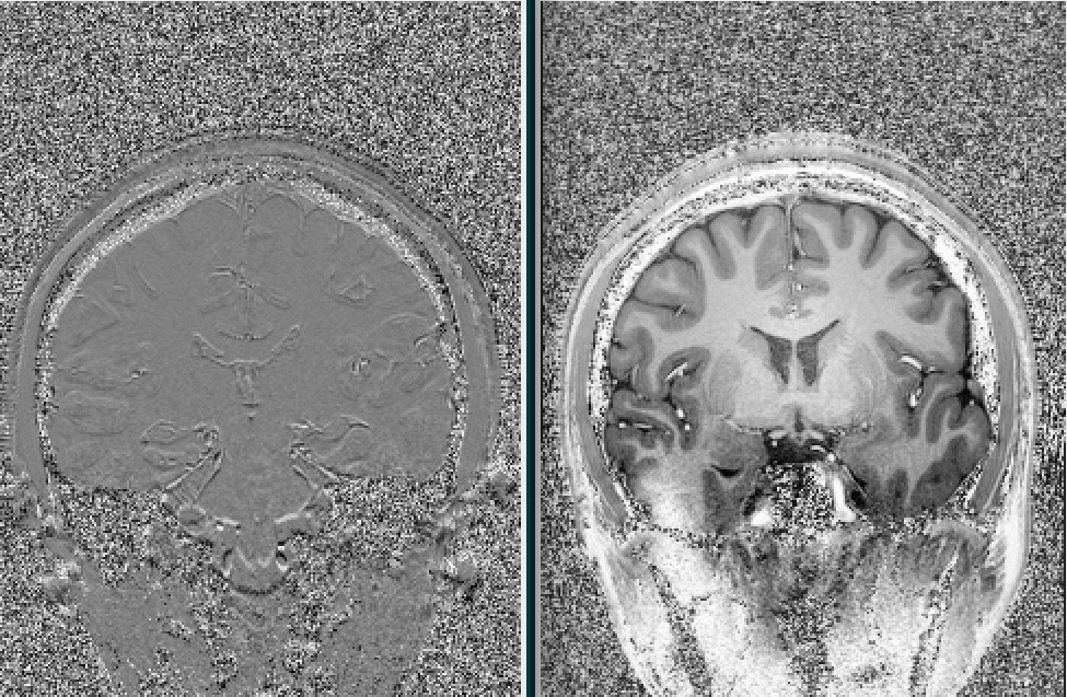
Figure1: MP2RAGE acquisition
Left: Difference image (odd -even echo). Image distortions induce signal enhancements/attenuations at tissue boundaries.
Right: Corresponding animated GIF illustrating the image distortions in greater detail.
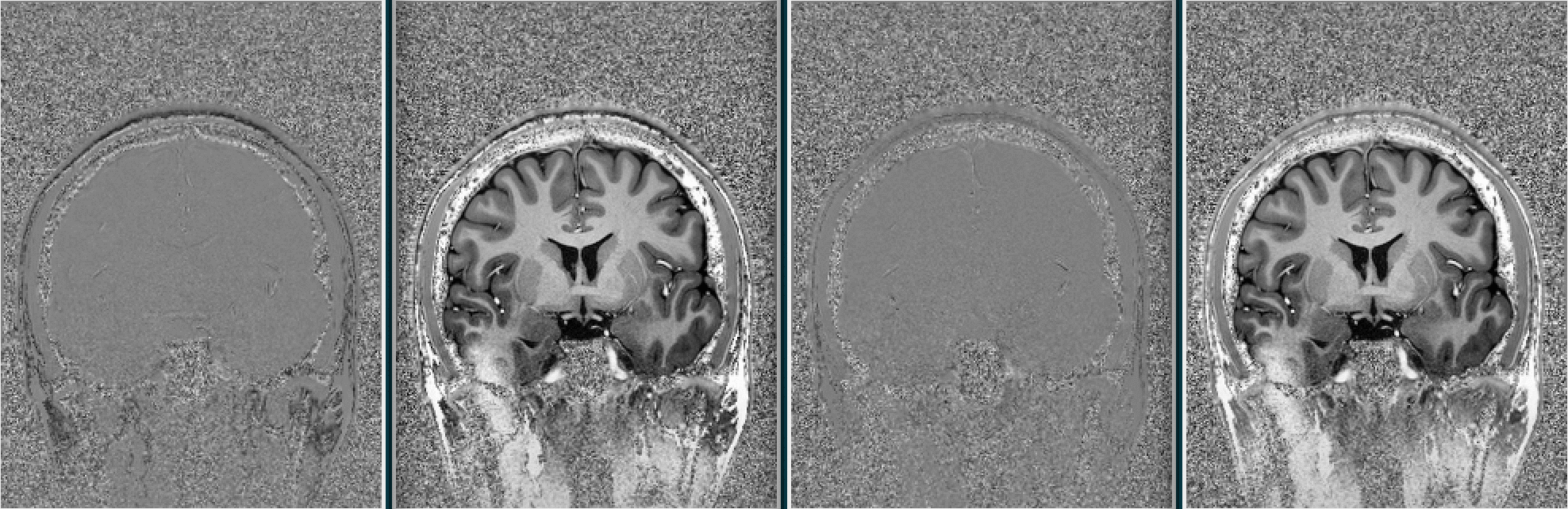
Figure 2: ME-MP2RAGE acquisition
Left: Composite image 1 (odd_odd-even_even echo). Image distortions are significantly reduced compared to the MP2RAGE acquisition.
Right: A further decrease in image distortions is achieved using composite image 2 (odd_even - odd_even echoes).
Animated GIF images are depicted as well for better illustration of locally induced image distortions.