1808
Implementation of the academic image processing pipeline ExploreASL in an outpatient center using IntelliSpace Discovery1Philips, Gainesville, FL, United States, 2Dent Neurologic Institute, Amherst, NY, United States, 3Helmholtz-Zentrum Dresden-Rossendorf, Dresden, Germany, 4Philips Research, Aachen, Germany, 5Dept of Radiology and Nuclear Medicine, Amsterdam University Medical Center, Amsterdam, Netherlands, 6Institute of Biomedical Engineering and Neurology, University College London, London, United Kingdom, 7Ghent Institute for Functional and Metabolic Imaging, Ghent, Belgium
Synopsis
The use of standardized image processing pipelines is continuously increasing in radiological research with developments in computing power, image processing, and machine learning techniques. Early integration of academic processing methods into clinical research workflow would accelerate the translation of promising novel MRI techniques into the clinic. However, the integration of such tools is both resource and time consuming. While most of neurological imaging takes place in outpatient centers, resource and workflow limitations of such clinics do not allow for the application of advanced image analysis. Here, we present the integration the “ExploreASL” into the PACS-connected research platform IntelliSpace Discovery.
Introduction
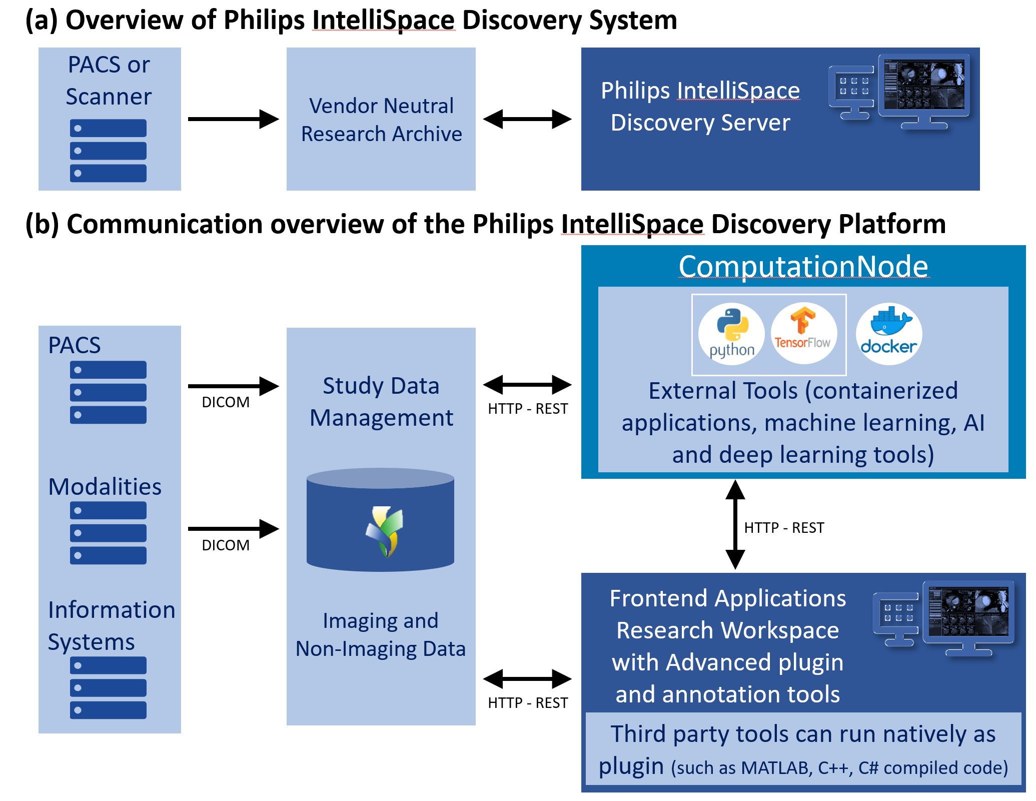
Novel academic image processing pipelines such as ExploreASL1 are not always accessible to clinical centers, because of a lack of resources or difficulty in integrating such tools for easy accessibility to radiologists. It remains challenging to bring such tools in the clinical radiology workflow for clinical validation. Here, we present the use of Philips IntelliSpace Discovery (ISD) (Philips Healthcare, Best, The Netherlands) for such purpose. ISD is a programmable platform that enables development, validation, and deployment of imaging processing assets into radiology infrastructure, with the aim to support clinical and translational research with advanced visualization and data management (Fig. 1a and 1b). We describe how such a platform can be used to deploy ExploreASL, a robust multi-vendor ASL image processing pipeline, at the Dent Neurologic Institute (DNI), a large outpatient center. ExploreASL encompasses a fully automated pipeline from import and structural image processing to cerebral blood flow (CBF) quantification and statistical analyses. ExploreASL is a clear example of a Matlab- and SPM-based research pipeline that, despite its standardization across different operating systems, MRI vendors and populations, it lacks the support for integration into a clinical workflow that is easy to use on a daily basis on a radiologist workstation. Therefore, we set out to implement ExploreASL as a plugin on the ISD server.Methods
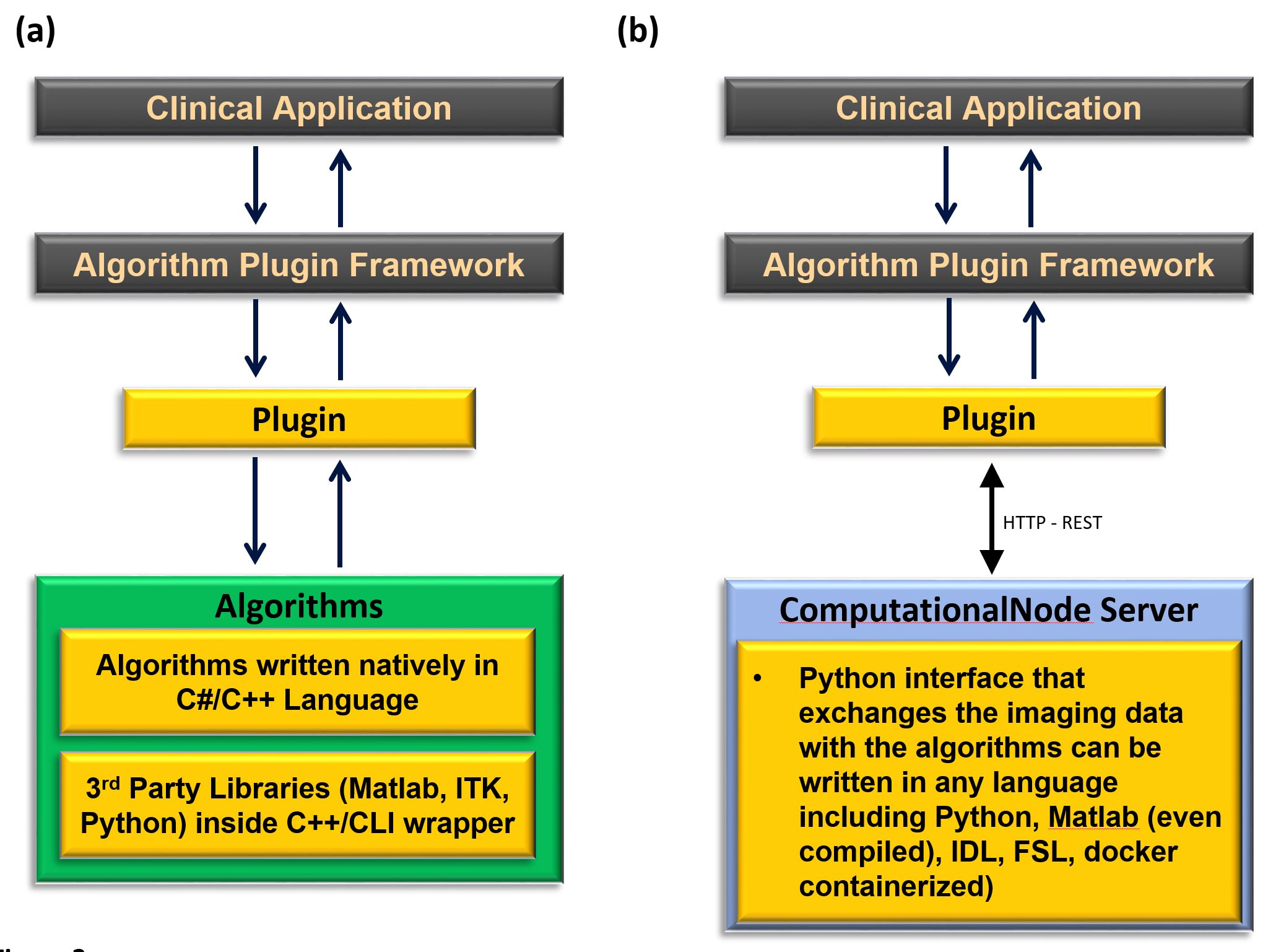
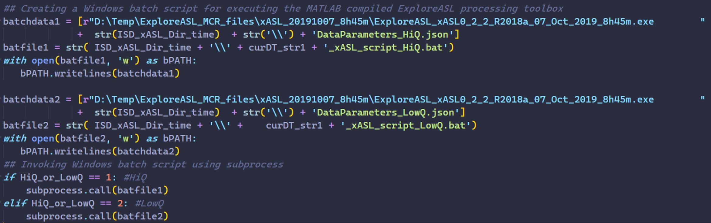
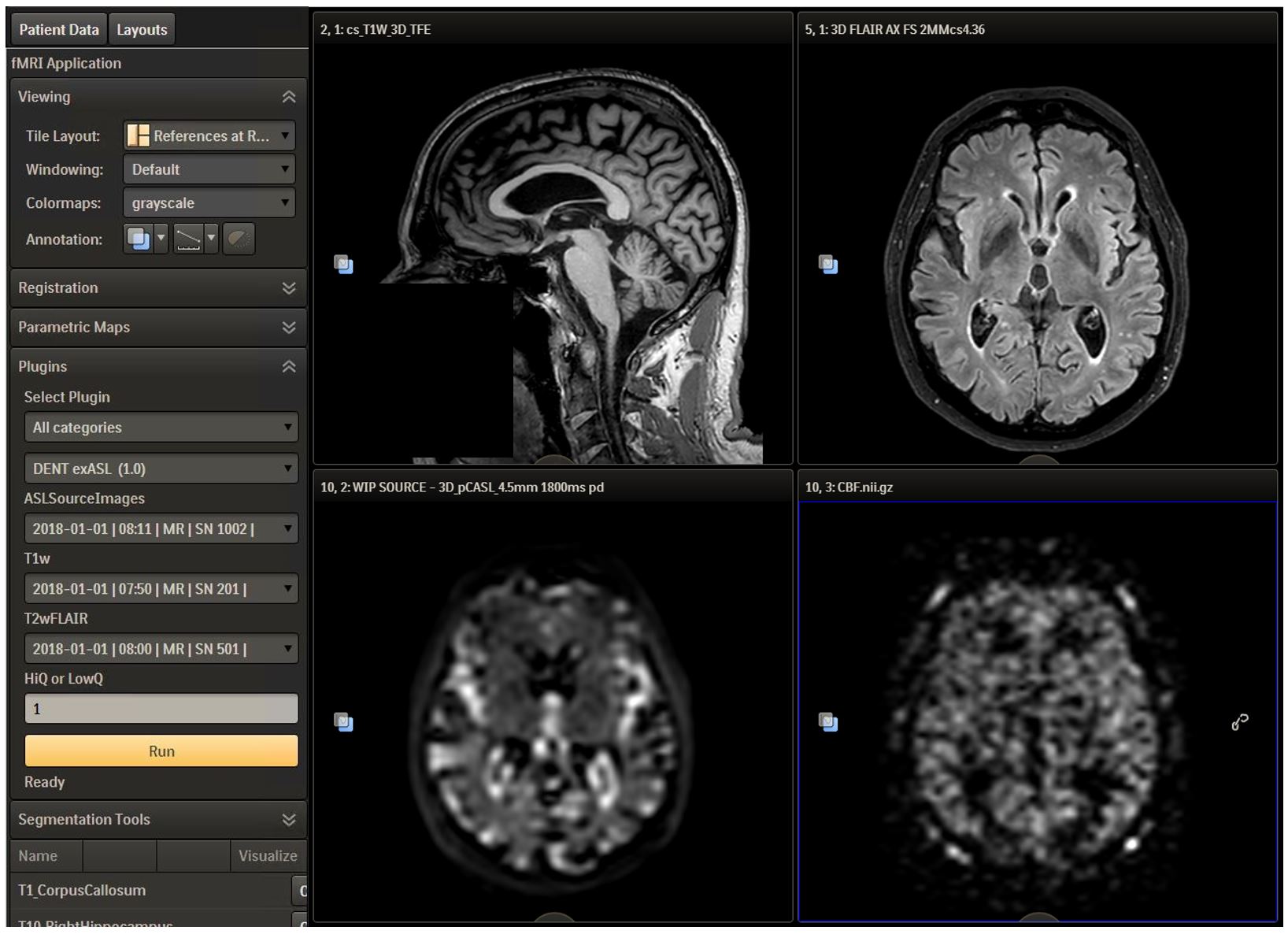
ExploreASL is written in MATLAB (MathWorks, MA, USA) and it is based on SPM122. To avoid implementing the entire ExploreASL pipeline as a native C# plugin, which would be very laborious and redundant work (Fig. 2a) in ISD, we used the ComputationalNode feature of the ISD, which allows a secure transfer of healthcare data (HTTP-REST standard) between ISD and external resources within a customizable Python wrapper (Fig. 2b).This ComputationalNode can be hosted either locally, or on a different server and/or different network, allowing for scalability of computation power. We designed an ISD-plugin for ExploreASL that takes the MRI data and converts them to BIDS3 compatible data formats, runs the compiled ExploreASL code (Fig 3.), and reads the output images and imports them back into the patient database. The wrapper takes 3D T1w, 3D FLAIR and 3D pseudocontinuous ASL (pCASL) as selected in the GUI (Fig. 4). As an initial implementation test, we used clinical MRI data of 14 patients (8f, 6m, age 74 ±5.9y) diagnosed with mild cognitive impairment, acquired at the DNI dementia clinic. To compare the performance and clinical utility of the plugin deployment, the images were processed with ExploreASL separately on a Windows laptop and on the ISD server with the deployed ISD plugin. The performance was validated qualitatively by a neuroradiologist with >10 years of experience (NP).Results
After the deployment of the ExploreASL on ISD, the interface allowed radiologists to initiate the processing of patient data and review the results from a web interface (Fig. 4). After image processing was performed, the generated quantified CBF maps were imported into the patient’s image dataset to allow visualization along with the structural images. Based on the 14 patients included in this pilot analysis, a qualitative comparison did not reveal major visual differences in lesion and GM-WM segmentation and CBF maps between the native and deployed version of ExploreASL. The average processing time per participant was 32:37 ± 4:50 and 48:40 ± 5:43 minutes, on a modern laptop (directly on Matlab) and with the ISD plugin, respectively, fitting with the processors being either optimized for single- or multi-threading. However, with parallel processing of data from seven subjects, the average processing time per participant dropped to 6:50 minutes on the ISD based analysis.Conclusion
We demonstrated the use of a research pipeline to analyze anatomical and ASL images directly on a clinical workstation, by a radiologist without prior experience or above-average IT skills. This allowed to overcome the technical and resource obstacles related to image processing, and enabled ASL quantification to be executed in the typical workflow of an outpatient practice. In the future, this would enable us to combine other existing plugins on the ISD server with the ExploreASL outputs to delineate regional changes in CBF. However, this needs to be validated. This approach creates the opportunity for non-academic centers to participate in clinical science and multicenter studies using ASL, which can facilitate the clinical validation of ASL and its incorporation into routine neuroradiology practice, as well as providing access to a large untapped source of data. This can also serve as a general model for creating clinically feasible image processing plugins to enable faster clinical validation and implementation of physiological MRI techniques.Acknowledgements
This work was supported by the Dent Family Foundation. HM is supported by AMYPAD grant no. 115952 and Amsterdam Neuroscience funding. FB is supported by NIHR funding through the UCLH Biomedical Research Centre.References
[1] Mutsaerts, H. J., Petr, J., Groot, P.. et al. ExploreASL: a collaborative effort to process and explore multi-center ASL data. Proceedings of the ISMRM 27th Annual Meeting & Exhibition (Vol. 27, p. 2705). International Society for Magnetic Resonance in Medicine (2019)
[2] Ashburner, J. SPM: A history. Neuroimage 62, 791–800 (2012).
[3] Gorgolewski, K., Auer, T., Calhoun, V. et al. The brain imaging data structure, a format for organizing and describing outputs of neuroimaging experiments. Scientific Data 3, 160044 (2016)
Figures