1789
Distortion Correction in EPI Diffusion MRI within Clinical Workflow: A Quantitative Evaluation1GE Healthcare, New York, NY, United States, 2Mount Sinai, New York, NY, United States, 3GE Healthcare, Waukesha, WI, United States, 4GE Healthcare, Menlo Park, CA, United States
Synopsis
This work does a quantitative evaluation of the effectiveness of a distortion correction method, clinically available at the scanner console, in diffusion MRI of the brain (N=10). After distortion correction, average cross-correlation coefficient between T1 and DWI was significantly increased. Distortion correction reduced average residual distance error to 1.1 mm or less (average 76% reduction) in all of three anatomical landmarks (frontal pole,lateral ventricle, pons). This on-console distortion correction method could help in the clinical applications that requires fast and accurate distortion correction such as deep brain stimulations surgery and cases with high susceptibility such as postoperative cohorts.
Introduction
EPI based diffusion MRI has been adopted widely in clinical brain MR exams but suffers from geometric distortions due to field inhomogeneities as well as eddy current. While diffusion distortion correction algorithms are routinely applied in research settings1, lack of a streamlined on-console processing and slow processing time limit their clinical adoption. Recently an advanced diffusion distortion correction which estimates the displacement map based on one additional reversed polarity gradient acquisition and outputs distortion corrected images automatically in the MR image browser, PROGRES (Polarity Reversed on Gradients for Reduced Susceptibility), was introduced as a clinical offering2. This work does a quantitative evaluation of the effectiveness of a distortion correction method in diffusion MR images of the brain.Methods
Data Acquisition: Ten healthy volunteers (age 46-75 years; mean age 59.1; 6F) were scanned under an IRB-approved protocol on a GE Premier 3T scanner (GE Healthcare, Waukesha, WI) using 48 channel head coil. Structural images were acquired using a 3D T1 MPRAGE with inhomogeneity correction (PURE) (146 slices, 1x1x1 mm3, TR = 2450 ms, TE = 2.94 ms, FA=8°). DWI datasets were acquired (3 directions, b=1000 s/mm2) using a single-shot SE-EPI sequence with distortion correction (PROGRES) and inhomogeneity correction (PURE) (FOV: 24 cm, matrix: 128 x 224, TR = 3952 ms, TE = 57.9 ms, slice: 32x4 mm, acceleration: 2 in-plane x 1 slice, NEX: 2, scan time: 1:06 min). Uncorrected DWI datasets were also automatically generated in the MR image browser.Processing: Data processing and visualization was accomplished using a combination of tools from AFNI (https://afni.nimh.nih.gov), FSL (https://fsl.fmrib.ox.ac.uk), MRtrix3 (https://www.mrtrix.org) package. The T1 structural image was skull-stripped and registered to native DWI space using a rigid registration tool. Affine registration matrix from T1 to MNI was also calculated. Contour of DWI was generated using the AFNI 3dedge3 program3 after resampling DWI to 0.5 mm isotropic resolution.
Quantitative Evaluation of Distortion:
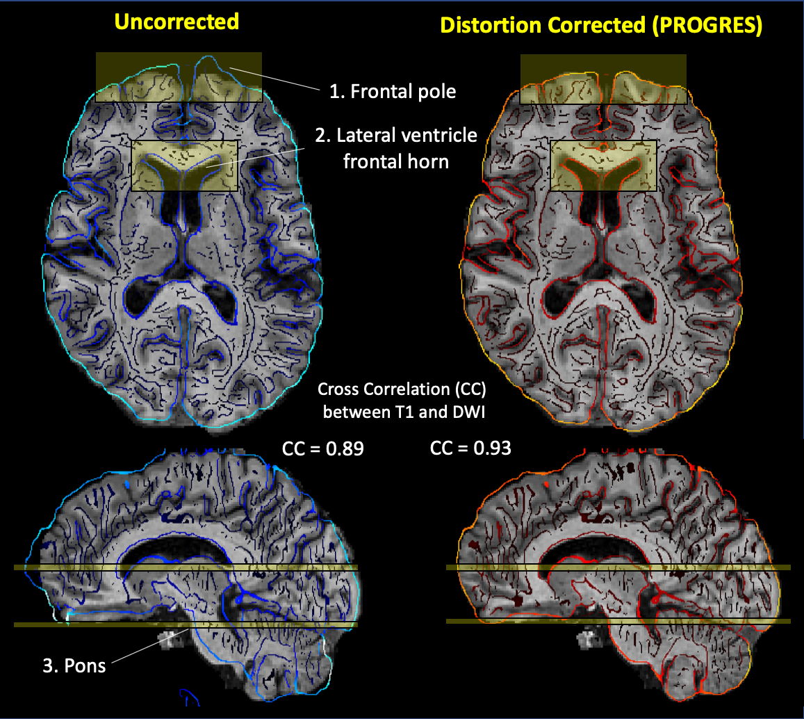
1) Cross-correlation between T1 and DWI, as a global similarity measure, was calculated before and after distortion correction. Then, Fisher’s z-transformation was applied before group comparison.
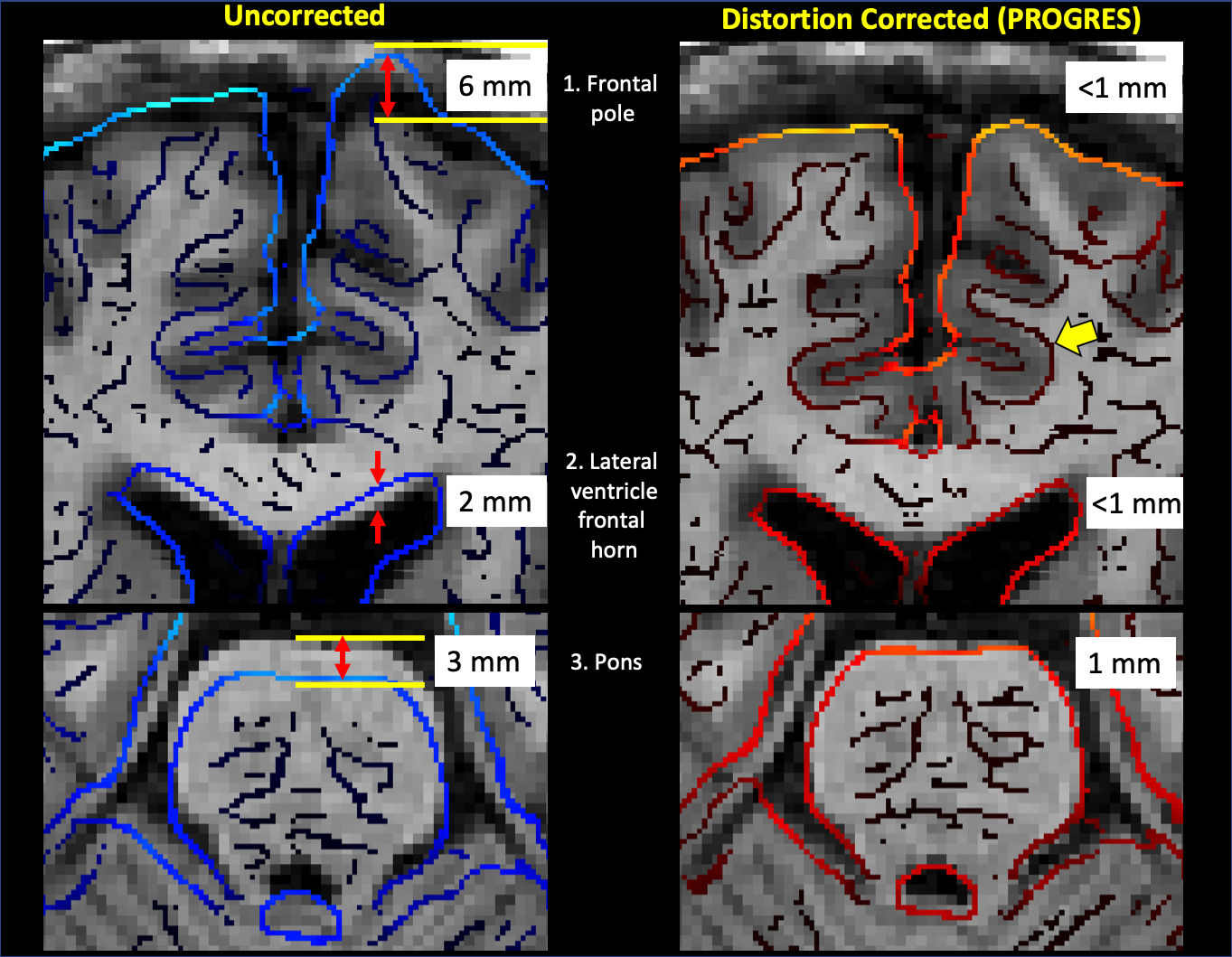
2) Residual distance error was measured by manually counting the number of voxels (voxel size = 1mm) in the distorted areas based on contour of DWI and co-registered T1 in three anatomical landmark (frontal pole, lateral ventricle frontal horn, pons) where the boundaries are clearly defined in T1 to avoid measurement errors and/or biasness (Figure 2).
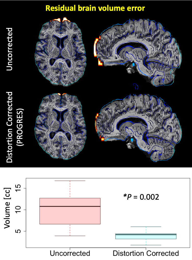
3) Residual brain volume error in frontal pole was calculated by automatically counting the number of voxels in the binary mask defined by the intersection of the following three masks: a) outside of T1 brain mask b) inside of DWI brain mask c) inside of mask in MNI coordinate (-30 < x < 30, y > 40, z < 30) registered to DWI native space via T1) .
Statistics: A two-sample t-test was used to compare two groups and a threshold of P< 0.05 (two-tailed) defined statistical significance.
Results and Discussion
A visual examination of the superimposed contours clearly reveals that distortion correction noticeably improved match of cortical and subcortical structures (Figure 1-2) including frontal pole, lateral ventricle frontal horn, brainstem and improved match of the boundary between white matter and grey matter (yellow arrow in Figure 2, in particular). This observation is consistent with the increase in a global similarity measure (from CC=0.89 to CC=0.93) (Figure 1). Average cross-correlation coefficient (N=10) was significantly increased from 0.87 to 0.92 (P= 3.8 x 10⁻⁹) (Figure 3).To quantify the extent of localized geometric distortion, residual distance error was measured in three anatomical landmarks (frontal pole, lateral ventricle, pons) (Figure 2). Local residual distance error in frontal role was reduced from 7.3 mm to 1.1 mm on average. In all of three anatomical landmarks, average residual distance error was significantly reduced to 1.1 mm or less (average 76% reduction) (Figure 3). Residual brain volume error (orange) in frontal pole significantly reduced with distortion correction from 10.12 cc to 3.92 cc (P= 0.002) (Figure 4).
Distortion correction method (PROGRES), which is clinically available at the scanner console, could be useful for the clinical applications that requires fast and accurate distortion correction such as the surgical targeting and postoperative evaluation of electrode location in deep brain stimulation surgery using diffusion weighted images and tractography4 as well as cases with high susceptibility such as postoperative cohorts and treatment/surgical planning5.
Acknowledgements
No acknowledgement found.References
1. Andersson, J. L., Skare, S., & Ashburner, J. (2003). How to correct susceptibility distortions in spin-echo echo-planar images: application to diffusion tensor imaging. Neuroimage, 20(2), 870-888.
2. Holland, D., Kuperman, J. M., & Dale, A. M. (2010). Efficient correction of inhomogeneous static magnetic field-induced distortion in Echo Planar Imaging. Neuroimage, 50(1), 175-183.
3. Monga, O., Deriche, R., Malandain, G., & Cocquerez, J. P. (1991). Recursive filtering and edge tracking: two primary tools for 3D edge detection. Image and vision computing, 9(4), 203-214.
4. Riva-Posse, P., Choi, K. S., Holtzheimer, P. E., McIntyre, C. C., Gross, R. E., Chaturvedi, A., ... & Mayberg, H. S. (2014). Defining critical white matter pathways mediating successful subcallosal cingulate deep brain stimulation for treatment-resistant depression. Biological psychiatry, 76(12), 963-969.
5. Treiber, J. M., White, N. S., Steed, T. C., Bartsch, H., Holland, D., Farid, N., ... & Chen, C. C. (2016). Characterization and correction of geometric distortions in 814 diffusion weighted images. PloS one, 11(3), e0152472.
Figures