1772
The effect of the refocusing flip angle on CSF dynamics imaging using multi-spin echo acquisition cine imaging (MUSACI)
Tatsuhiro Wada1, Chiaki Tokunaga1, Osamu Togao2, Yasuo Yamashita1, Kouji Kobayashi1, Masami Yoneyama3, and Toyoyuki Kato1
1Division of Radiology, Department of Medical Technology, Kyushu University Hospital, Fukuoka, Japan, 2Department of Clinical Radiology, Graduate School of Medical Sciences, Kyushu University, Fukuoka, Japan, 3Philips Japan, Fukuoka, Japan
1Division of Radiology, Department of Medical Technology, Kyushu University Hospital, Fukuoka, Japan, 2Department of Clinical Radiology, Graduate School of Medical Sciences, Kyushu University, Fukuoka, Japan, 3Philips Japan, Fukuoka, Japan
Synopsis
Multi-spin echo acquisition cine imaging (MUSACI) is based on the multi-spin echo technique and used for detection of cerebrospinal fluid (CSF) movement. MUSACI detects CSF movement as a signal loss due to proton phase dispersion and flow void. To better detect the CSF loss caused by CSF movement and reduce the CSF signal loss caused by T2 decay, we modified the refocusing flip angle modulation using pseudo steady state sequence (PSS). The modulation of refocusing flip angle in PSS improved the CSF dynamic imaging using MUSACI.
INTRODUCTION
Hydrocephalus is an abnormality of the cerebrospinal fluid (CSF) circulation caused by aqueductal stenosis, tumors, etc. A novel magnetic resonance (MR) imaging technique, multi-spin echo acquisition cine imaging (MUSACI), has been used to describe the movement of the CSF.1 This technique is based on the proton phase dispersion and flow void using 3D multi-spin echo imaging. However, in the 3D turbo spin-echo (TSE) sequence, CSF signal loss can also occur when the refocusing flip angle modulation is low.2 The purpose of this study was to investigate the effect of modifying the refocusing flip angle on the CSF dynamic imaging using MUSACI.METHODS
The MUSACI images were acquired in 10 healthy volunteers (7 men and 3 women; age range 24–44 years; mean age 29.4 ± 6.2 years). All images were acquired on a 3T MR scanner (Ingenia, Philips, Best, The Netherlands) using a 15-channel head coil. MUSACI images were acquired in the sagittal plane using 3D VISTA with pulse gating. The parameters were TR = 2RR msec, TE = 44.0 + echo number (80 msec steps) msec, TSE factor = 70–110, echoes = 7–10, field of view = 240 × 240 mm2, slice thickness = 1.2 (recon to 0.6) mm, number of slices = 25, matrix size = 416 × 416, scan time = approx. 5 min, refocusing flip angle = constant 30°, constant 50°, constant 80°, pseudo steady state (PSS) 50°–70°–100° (PSS 50°), PSS 80°–100°–130° (PSS 80°) (Fig. 1). We modified the TSE factor and echo numbers so that the TR was within one pulse wave in each study. In this study, circular regions of interest were placed to measure the signal intensity (SI) in the lateral ventricle, the foramen of Monro, the third ventricle, the fourth ventricle and the pons in MUSACI. The Pearson’s correlation coefficients were calculated to evaluate the statistical correlation between the CSF SI and TEeff in the lateral ventricle. Values of p<0.05 were considered significant.RESULTS
Figure 2 shows the influence of refocusing flip angle modulation on the midsagittal MUSACI images in a 26-year-old female. The antegrade and retrograde directions of CSF movement were detected in all sequences. The CSF signal loss caused by CSF movement was greater when the refocusing flip angle was lower under both the constant and PSS sequences. Figure 3 shows the influence of refocusing flip angle modulation on the foramen of Monro MUSACI images in a 26-year-old female. The antegrade direction of CSF movement was detected in all constant sequences. In the PSS sequences, on the other hand, it was detected in PSS 50° but was not clearly detected in PSS 80°. The CSF signal loss caused by CSF movement was more apparent than the CSF signal decay by the lower refocusing flip angle under both the constant and PSS sequences. Figure 4 shows the SI change with an increase in TEeff in each sequence. The reductions of the CSF SI in the foramen of Monro, third ventricle and fourth ventricle were steeper than that in the lateral ventricle. The CSF SI was decreased when using the lower refocusing flip angle under both the sequence constant and PSS sequence conditions, and those under the PSS sequence were higher than those under the constant sequence conditions. Figure 5 shows the change of CSF SI in the lateral ventricle with an increase in TEeff using MUSACI at all sequences. A severe reverse correlation between the CSF SI and TEeff was observed at constant 30° (r=-0.960, p<0.0001), 50° (r=-0.971, p<0.0001) and 80° (r=-0.875, p=0.0009), a weak positive correlation between the CSF SI and TEeff was observed at PSS 50° (r=0.280, p=0.4340), and a moderate reverse correlation between the CSF SI and TEeff was observed at PSS 80° (r=-0.597, p=0.0683).DISCUSSION
The CSF signal loss caused by CSF movement in MUSACI was more apparent when using a lower refocusing flip angle than a higher refocusing flip angle. The sensitivity of the refocusing flip angle on flow using fast-spin-echo sequence has been reported.3,4 The magnetization at each echo in turbo-spin echo is no longer rephased completely into the transverse plane when the refocusing flip angles are less than 180°. The phase dispersion caused by moving protons was accelerated due to a detour in the longitudinal signal pathways when using a low refocusing flip angle. The relationship between the CSF signal and TEeff exhibited a reverse correlation in the constant 30°, 50°, 80° and PSS 80° sequences, but a positive correlation was observed in the PSS 50° sequence. The PSS sequence yielded a higher echo signal of CSF than the constant sequence due to the use of the variable flip angle.5,6 In addition, a lower refocusing flip angle could maintain the signal variation of CSF to the level of the constant in the PSS sequence.CONCLUSION
The CSF dynamic imaging using MUSACI was improved by modifying the refocusing flip angle. We selected PSS 50° sequence as the optimal parameter, because the CSF signal loss due to CSF movement was changed by modulating the flip angles of the initial refocusing RF pulses and the use of a variable refocusing flip angle also reduced the CSF signal decrease caused by T2 decay.Acknowledgements
No acknowledgement found.References
- Wada T, Tokunaga C, Togao O, et al. Visualization of cerebrospinal fluid dynamics using multi-spin echo acquisition cine imaging (MUSACI). Magn Reson Med 2019;81(1):331-341.
- Busse RF. Flow Sensitivity of CPMG Sequences with Variable Flip Refocusing and Implications for CSF Signal Uniformity in 3D-FSE Imaging. In: Proceedings of the ISMRM, Seattle; 2006. p.2430.
- Yoneyama M, Nakamura M, Tabuchi T, Takemura A, Obara M. Optimization of 3D-variable refocusing flip angle RARE imaging for high-resolution volumetric black-blood angiography. Radiol Phys Technol 2012;5(2):270-276.
- Storey P, Atanasova IP, Lim RP, et al. Tailoring the flow sensitivity of fast spin-echo sequences for noncontrast peripheral MR angiography. Magn Reson Med 2010;64(4):1098-1108.
- Busse RF, Brau AC, Vu A, et al. Effects of refocusing flip angle modulation and view ordering in 3D fast spin echo. Magn Reson Med 2008;60(3):640-649.
- Mugler JP, 3rd. Optimized three-dimensional fast-spin-echo MRI. J Magn Reson Imaging 2014;39(4):745-767.
Figures
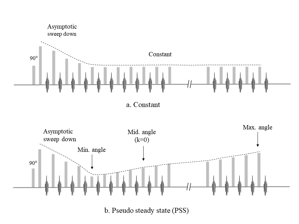
Fig 1. The schema of refocusing flip angle modulation constant (a) and pseudo
steady state (PSS) (b).
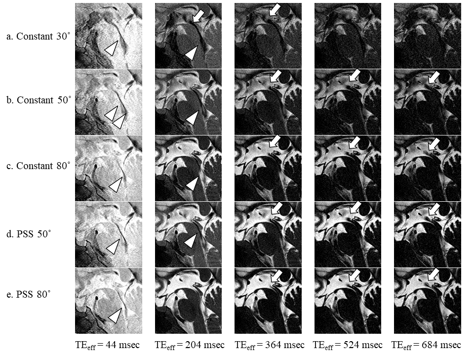
Fig 2.
Midsagittal brain MUSACI images in a healthy volunteer. The anterograde CSF
movement from the aqueduct to the fourth ventricle (arrow heads), and the
retrograde movement from the aqueduct to the third ventricle (arrows) are
observed in all sequences.
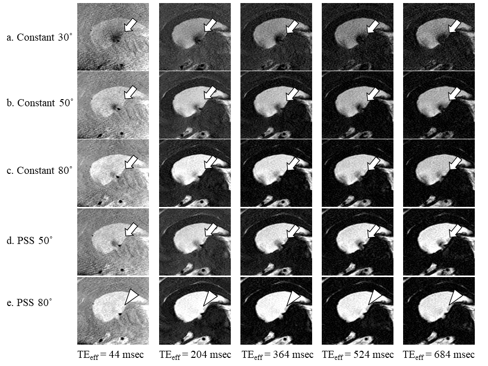
Fig 3. Sagittal
brain MUSACI images at the section of the foramen of Monro in a healthy
volunteer. The retrograde CSF flow from the foramen of Monro to the lateral
ventricle is observed on the images of all constant sequences (a-c) and PSS 50°
(d) (arrows); however, it was not clearly detected in PSS 80° (e) (arrow
heads).
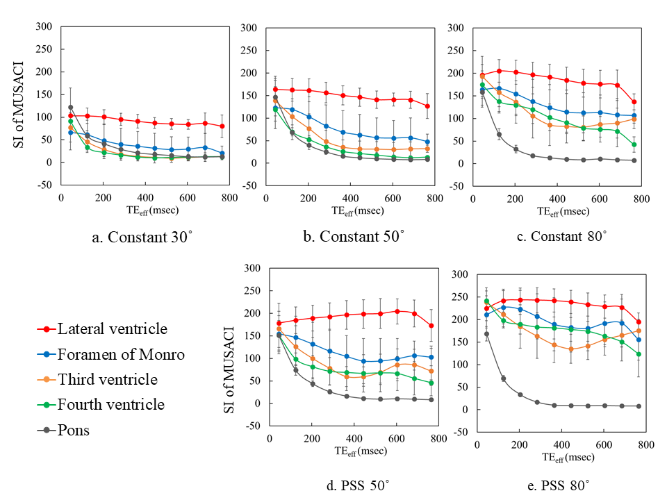
Fig 4. The SI change with increase in TEeff in constant 30° (a),
constant 50° (b), constant 80° (c), PSS 50° (d), PSS 80° (e). The CSF SI was
decreased when using a lower refocusing flip angle in both sequence constant
and PSS.
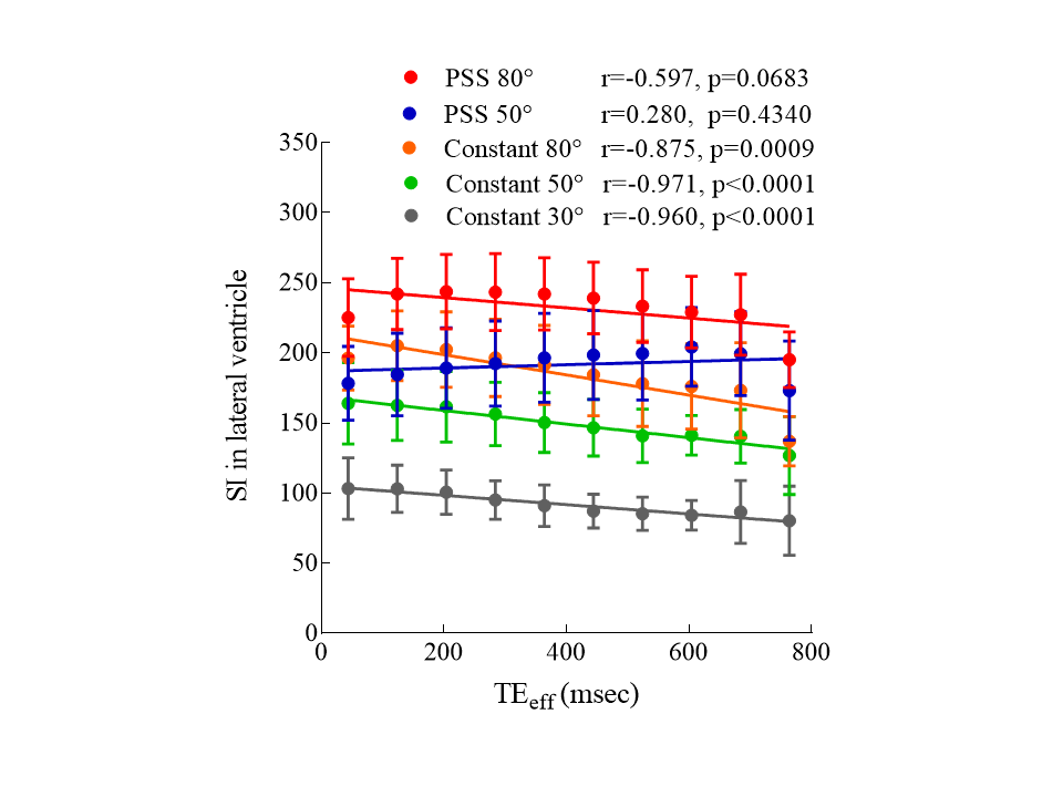
Fig 5. The change of CSF SI in the lateral ventricle with an increase in TEeff
using MUSACI at all sequences. A severe reverse correlation between the CSF SI
and TEeff was observed at constant 30° (a), 50° (b) and 80° (c), a
weak positive correlation was observed between the CSF SI and TEeff
at PSS 50° (d), and a moderate reverse correlation was observed between the CSF
SI and TEeff at PSS 80° (e).