1769
Combination of Compressed Sensing and Sensitivity Encoding (CS-SENSE) in MR brachial plexus imaging: a study of different acceleration factors1radiology, Union Hospital, Tongji Medical College, Huazhong University of Science and Technology, Wuhan, China, 2Clinical Science, Philips Healthcare, Shenzhen, China, 3Clinical Science, Philips Healthcare, Beijing, China, 4Philips Healthcare, Beijing, China
Synopsis
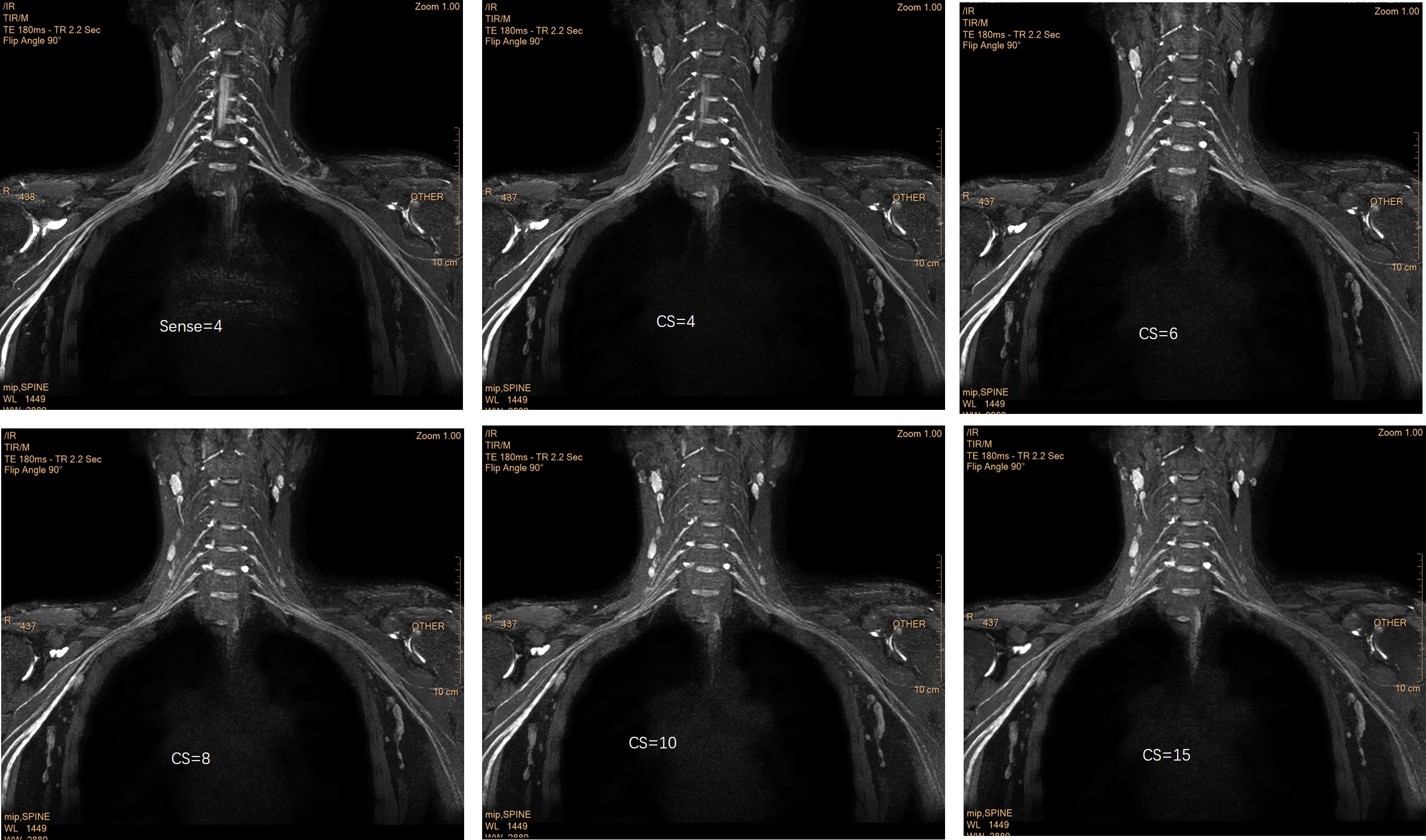
The aim of this study was to reduce the scan time of 3D Nerve-view using Compressed Sensing-Sensitivity Encoding (CS), and evaluate the image quality and capability of diagnosis of accelerated 3D Nerve-view sequences. 3D Nerve-view sequences with 5 different CS (compressed sense technology) accelerating factors (4,6,8,10,15), and a traditional 3D Nerve-view with 4-fold parallel imaging (sense) as a clinical reference were obtained.The 3D-CS sequence offer comparable diagnostic quality to the clinical 3D scan with much less time, potentially increasing the productivity of MR scanners.CS-3D Nervview with factor 6 offer equilibrium between comparable clinical diagnostic quality with less scan time (235seconds).
Objective
High resolution visualization of brachial plexus using 3D Turbo-Spin-Echo (3D-TSE) sequence with contrast-enhancement is important for the evaluation of brachial plexus diseases(1). Motion artifacts have become challenging to image quality with increasing three-dimensional resolution due to the increasing imaging time(2). The objective of this study was to accelerate 3D-TSE-based brachial plexus imaging with a combination of compressed-sensing and sensitivity-encoding (CS-SENSE) techniques and evaluate the image quality and diagnosis performance at different acceleration factors.Methods
Forty-five patients with suspected brachial plexus diseases were consecutively enrolled, who underwent MR studies on a 3T scanner (Ingenia CX, Philips Healthcare, Best, the Netherlands), and study approved by the local IRB. 3D-TSE sequences (commercially available as 3D-NerveView) with 5 different CS-SENSE acceleration factors (4,6,8,10,15) were scanned and compared against the same sequence with 4-fold SENSE acceleration (clinical reference). All other imaging parameters were kept the same. All images were graded by two radiologists with >10 years’ experience in MR neurography for image quality (on a 5-point scale). Quantitative analysis was performed to evaluate the image quality on signal-to-noise ratio (SNR) and contrast-to-noise ratio (CNR). The similarity between the CS-SENSE accelerated images and the clinical reference was evaluated using the pixelwise root mean square error (RMSE) and structural similarity index (SSIM) (3).The scan time of each sequence were recorded. An analysis of variance with repeated measurements and the Friedman test was used to test for potential difference between the sequences.Results
The mean values of the RMSE ranged from 73.38 ± 15.91 for CS 4 to 234.66 ± 43.56 for CS 15, while SSIM was highest for CS 4 with 99.21% ± 2.23% and lowest for CS 15 with 87.90% ± 5.32%. The scan time with SENSE 4,CS-SENSE 4, CS-SENSE 6, CS-SENSE 8, CS-SENSE 10, and CS-SENSE 15 was 669s,350s,235s,176s,143s,95s, respectively. The mean subjective score from both radiologists was 4.2 0.3 for CS-SENSE images with acceleration rate below 8. There was no statistical difference in the brachial plexus to surrounding tissue contrast between CS factor 4-6, and the lesion display of the brachial plexus had no statistical difference. The images of CS factor above 10 had no diagnosis value, implying a maximum CS-SENSE acceleration factor of 6 in this study.Conclusion
In conclusion, 3D-TSE contrast-enhanced sequence with CS-SENSE acceleration rate up to 6 with satisfactory diagnostic performance and dramatically reduced imaging time (reduced by 64.9% from the clinical reference), which may potentially enhance the hospital workflow.Acknowledgements
Many thanks to Department of Radiology, Union Hospital, Tongji Medical College, Huazhong University of Science and Technology.
Many thanks to MR Collaborations, Clinical Science, Philips Healthcare, China
References
1. Wang L, Niu Y, Kong X, et al. The application of paramagnetic contrast-based T2 effect to 3D heavily T2W high-resolution MR imaging of the brachial plexus and its branches. Eur J Radiol 2016; 85:578-584.
2. Klupp E, Cervantes B, Sollmann N, et al. Improved Brachial Plexus Visualization Using an Adiabatic iMSDE-Prepared STIR 3D TSE. Clin Neuroradiol 2018.
3. Vranic JE, Cross NM, Wang Y, Hippe DS, de Weerdt E, Mossa-Basha M. Compressed Sensing–Sensitivity Encoding (CS-SENSE) Accelerated Brain Imaging: Reduced Scan Time without Reduced Image Quality. Am J Neuroradiol 2019; 40:92-98.
Figures