1754
In vivo performance evaluation of silicon carbide dielectric pads for 7T MRI.1Multiwave Imaging, Marseille, France, 2Aix Marseille Université, Marseille, France, 3CEA-DRF/Joliot/Neurospin, Gif-sur-Yvette, France, 4CEA-DAM Le Ripault, Monts, France, 5CEA-DRF/IRAMIS/NIMBE/LSDRM, Gif-sur-Yvette, France, 6Faculty of Physics and Engineering, ITMO University, Saint Petersburg, Russian Federation
Synopsis
Dielectric pads have demonstrated to be a simple and yet efficient solution to mitigate locally RF transmission heterogeneities while using classic 7T MRI birdcage. Extensive research has not yet been carried on alternative candidate to water-based perovskites. The grail would be a long-lasting, comfortable, MR invisible, efficient, unalterable and high permittivity soft material. In this study, a novel material based on silicon carbide particles and addressing those requirements was successfully compared to perovskite pads from the literature in terms of B1+ homogeneity and image contrast through in vivo measurements.
Background
With higher signal and contrast-to-noise ratio, MR neuroimaging at UHF (≥ 7 Tesla) promises remarkable improvements in spatial and/or temporal resolutions. However, several limitations have been reported, hindering the realization of these promises for clinical research. The main issue is the B1+ field inhomogeneities causing variable excitation of the nuclear spins across the brain dielectric. This results in signal void areas or contrast losses in the subsequent MR images, rendering them unexploitable for any potential diagnosis. One simple, yet efficient way to address those inhomogeneities is to install pads made of High-Dielectric Constant (HDC) materials based on perovskites and heavy water inside a volume birdcage coil, classically used for RF transmission at UHF [1]. While HDC perovskite-based pads are already available on the market [2], we are proposing a new improved design for HDC pads based on silicon carbide particles optimized with regard to permittivity, size and SAR [3-4]. The present work aims at evaluating in vivo the performance of those new pads in terms of performance. Therefore, they are compared to state-of-the-art perovskites aqueous pads (namely BaTiO3 and CaTiO3) [5,6]. We demonstrate in the present work that our new pads efficacy is on pair with conventional pads and exhibit additional features such as MRI invisibility and long-term usability.Methods
Pads efficiency were experimentally tested on healthy volunteers on a 7T Magnetom scanner (Siemens Healthineers, Erlangen, Germany) with a single transmission RF-coil and a 32 phased array coil for reception (Nova Medical, Willington, MA, USA). B1+ maps and proton density low spatial resolution images were acquired through a clinical protocol designed to safely assess new MR RF prototypes [7]. The study was approved by the local ethics committee and volunteers gave written informed consent. Pads dimensions and materials are presented in Table 1. To investigate the potential chemical shift artefacts from the solvent and dispersant hydrogen bonds, SiC pad mixture T2 and T2* relaxation times were characterized at 7T in NMR spectrometer (Brucker, Ettlingen, Germany) with non-selective MR spectroscopic Multi-Slice-Multi-Echo (MSME) (7×TE with ΔTE 5.2ms) and Free Induction Decay (FID) sequences respectively.Results
B1+ field distributions obtained on one volunteer (Figure 1) showed that the B1+ mean field magnitudes in the temporal lobes increased up to 33% in the case of the CaTiO3 and SiC pads and up to 47% in the case of the BaTiO3 pads which lead to enhanced contrast-to-noise ratio on brain proton density images. It can be also noted that BaTiO3 pads are observed on the images. Although we have not conducted experiments, we believe this is due to deuterium atoms from the heavy water exchanging with the hydrogen atoms in the plastic pouch. In comparison, SiC pads remained invisible on the image. Further investigations on T2* and T2 relaxation times measured using FWHM of the FID and MSME signal decay respectively (Figure 2) showed extremely low values that made the pads invisible for most of the classic MR image weightings.Discussion
SiC pads meet the specifications set by the existing perovskites standards while featuring additional properties that outclass them. On the one hand, they robustly enhance the local B1+ field leading to an improved contrast in the temporal lobes. On the other hand, they remain invisible due to the nature of the solvent. In addition, overall cost is lowered as deuterated water is not involved and, finally, there are fewer restrictions in terms of preparation conditions during elaboration process and safety measures while used on a daily basis as the material is not harmful [9-10].Conclusion
We demonstrated that this novel material is a promising perovskite aqueous pads replacement candidate. To confirm those results and assess inter subjects variability, a study with more volunteers at full power needs to be carried out in the future.Acknowledgements
This work has been supported by the Programme
Transversal du CEA and the Leducq Foundation large
equipment ERPT program and the NEUROVASC7T project. Results incorporated in this work also received funding from the the FET-OPEN M-CUBE Project under grant agreement No #736937 and from FET-OPEN
MRI PADS launchpad Project under grant agreement No #850506.
References
[1] Webb AG, Dielectric materials in magnetic resonance, Concepts in Magnetic Resonance Part A, 2011;38A(4):148‑84.
[2] Multiwave Imaging: www.multiwaveimaging.com/products/mri
[3] Raolison Z, Dubois M, Neves AL, Enoch S, Mallejac N, Sabouroux P, Adenot-engelvin AL, Vignaud A, Abdeddaïm R, Properties optimization of pads configurations on CST, ISMRM 2018 Paris, France; 2018.
[4] Raolison Z, Abdeddaïm R, Leroi L, Neves AL, Dubois M, Mauconduit F, Luong M, Enoch S, Mallejac N, Sabouroux P, Adenot-Engelvin AL, Vignaud A, Evaluation of a new long-lasting silicon carbide based dielectric pad for ultra-high field MRI, ISMRM 2018 Paris, France; 2018.
[5] O’Reilly TPA, Webb AG, Brink WM, Practical improvements in the design of high permittivity pads for dielectric shimming in neuroimaging at 7T, J Magn Reson, 2016;270:108‑14.
[6] Neves AL, Leroi L, Raolison Z, Cochinaire N, Letertre T, Abdeddaïm R, et al., Compressed perovskite aqueous mixtures near their phase transitions show very high permittivities: New prospects for high-field MRI dielectric shimming, Magn Reson Med. 2018;79(3):1753‑65.
[7] Vignaud A, Mauconduit F, Gras V, Girard O, Kober F, Hertz-Pannier L, et al. Fast and unconditionally safe in vivo MR head protocol for home-made coil prototype assessment at 7T, In: Proceedings of the ESMRMB. Rotterdam, Netherlands; 2019.
[8] Haines K, Smith NB, Webb AG, New high dielectric constant materials for tailoring the B1+ distribution at high magnetic fields, J Magn Reson, 2010;203(2):323‑7.
[9] Barium Titanate Oxide: MSDS No. 12348. Alfa Aesar website. Accessed August 29, 2018.
[10] Fluoroethylene carbonate: MSDS No. 3205E-2. Kishida chemicals website. Accessed August 29, 2018.
Figures


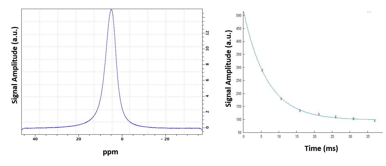
Figure 2 – On left plot, SiC mixture FID fast Fourier transform at 7T having a Full Width at Half Maximum (FWHM) Δν=1/π T2*=1700 Hz leading to T2*=0.19 ms. A gaussian fit was used directly on the FFT signal for searching the FWHM. On the right plot, same mixture analyzed with MSME leading to very short T2=6.6ms.