1747
Compressed Sensed MPRAGE with Parallel Imaging: Image Quality Metrics and Morphometry Study at 3T1GE Healthcare, Menlo Park, CA, United States, 2GE Healthcare, Rochester, MN, United States, 3GE Healthcare, Hino Tokyo, Japan
Synopsis
While there is a growing trend for using volumetric acquisitions for brain MRI, long acquisition times still limit their adoption. There is still a tremendous need to reduce scan time of these volumetric acquisitions to improve workflow productivity and to reduce the likelihood of motion during the scan. MPRAGE sequence is a key 3D brain acquisition because of its excellent gray-white contrast which is ideal for visualization of substructures, segmentation and morphometric measurements. This work takes a rigorous quantitative approach to investigating the effects of compressed sensing and parallel imaging factors on various tissue measurements derived from accelerated MPRAGE images.
INTRODUCTION
While there is a growing trend for using volumetric acquistions for brain MRI, long acquisition times still limit their adoption. There is still a tremendous need to reduce scan time of these volumetric acquisitions to improve workflow productivity and to reduce the likelihood of motion during the scan.IR prepared MPRAGE is an important sequence in neuroimaging as it provides excellent WM/GM contrast and a high spatial resolution. Consequently, the MPRAGE images are commonly used to segment brain tissues, derive morphometric measurements as well as to align datasets acquired at a lower resolution (e.g. fMRI, DTI) to a standard space (e.g. MNI, Talairach). However these scans can typically take 5-6 mins to acquire.
Parallel imaging and compressed sensed (CS) undersampling are two commonly applied techniques to reduce scan time in 3D imaging. In this study, we acquired MPRAGE images at varying levels of combined parallel imaging and CS undersampling and evaluated the dependence of increased acceleration factor on 1) the image quality and 2) the estimates of cortical thickness and cortical/subcortical volume. The overall objective was to find out how far acceleration can be pushed with the MPRAGE acquisition without compromising its utility.
METHODS
Three healthy volunteers (2 females) were scanned on a 3T MR750 Discovery scanner (GE Healthcare, Milwaukee, WI) with the 32-ch Nova head coil (Nova Medical, Wilmington, MA).MPRAGE data was acquired sagittally using the scan parameters recommended in the ABCD Study protocol (https://abcdstudy.org/scientists/protocols), i.e. TR/TE/TI=2500ms/2ms/1060ms, FA=8°, 256x256, FOV: 256mm2.
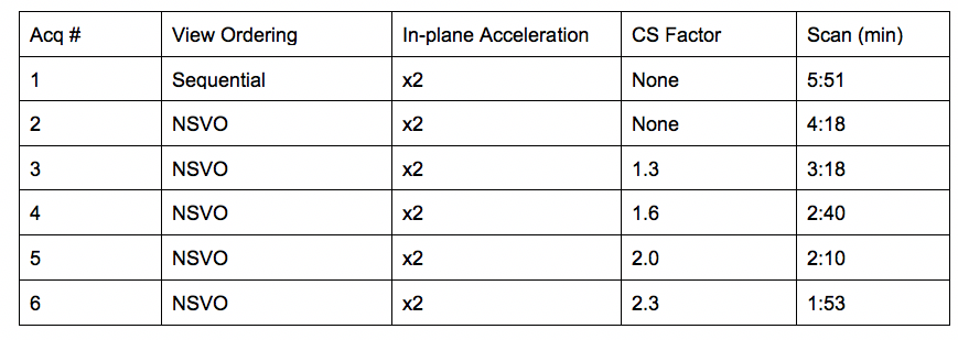
The MPRAGE scan was repeated at an increasing level of acceleration factor using the parameters listed in Table 1. The under sampled data were acquired using a segmented non-separable ky-kz view ordering (NSVO) scheme and were reconstructed using a CS+ARC algorithm described in reference 1 and 2.
All images were converted to BIDS data structure and validated (https://github.com/bids-standard/bids-validator). Subsequently, the data sets were fed into the MRIQC BIDS-App (v0.15.2rc1) to extract standard image quality metrics (https://mriqc.readthedocs.io/en/stable/index.html).
Additionally, all data sets were also processed through FreeSurfer (v6.0.0, http://surfer.nmr.mgh.harvard.edu on an AWS EC2 t2.2xlarge instance) to estimate the cortical/subcortical volumes.
RESULTS
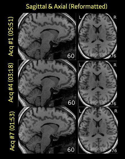
Figure 1 shows sagittal and reformatted axial images acquired at different acceleration levels (reference, mid, high) from a representative subject and as output by the MRIQC visual report.The overall image quality is well preserved even at the highest acceleration factor with more than 60% in time savings.
Figure 2 shows the MRIQC tissue-combined SNR metric plotted as a function of the increased acceleration factor for all subjects.
Figure 3 shows the percent change in FreeSurfer volume estimates relative to the reference scan (Acq #1) for cortical gray matter (A), ventricles (B), cerebral white matter (C), and subcortical gray matter (D).
DISCUSSION & CONCLUSION
Both in terms of quantitative and qualitative assessments, we found increased acceleration had no major impact on image quality and morphometric measures. Notably the SNR metric improved as acceleration increased. This is likely due to the regularization effect of the CS with parallel imaging reconstruction.Acknowledgements
No acknowledgement found.References
1. King et al, Proc. Intl. Soc. Mag. Reson. Med. 18 (2010), 4881
2. Anja, Brau et al, MRM 2008;59:382
Figures