1745
The difference between multicentric gliomas and diffuse gliomas: evidence from the probabilistic fiber tracking by diffusion tensor imaging
Simin Zhang1, Xiaorui Su1, Weina Wang1, Qiang Yue1, and Qiyong Gong1
1Huaxi MR Research Center,Department of Radiology, West China Hospital of Sichuan University, Chengdu, China
1Huaxi MR Research Center,Department of Radiology, West China Hospital of Sichuan University, Chengdu, China
Synopsis
Multicentric gliomas are rare lesions of central nervous system. Due to the invasive nature of glioma, differentiating multicentric gliomas from diffuse gliomas is difficult. As yet there remains lack of methods to elucidate lesions are totally separated unless surgical biopsy. We use probabilistic fiber tracking to identify the white matter dissemination routes. Our results showed the connectivity value and probabilistic value of the lesions in multicentric gliomas were significantly lower than the lesions in diffuse gliomas. The features provide new insight into multicentric gliomas and may aid in accurate classification of multicentric glioma and diffuse glioma in vivo.
Object
Multicentric gliomas are uncommon lesions of the central nervous system. Till now, the evolution of multicentric gliomas is poorly understood. Due to the white matter infiltration of glioma, it is hard to exclude the existence of a connection in cases deemed to be multicentric. As yet there remains lack of methods to elucidate the lesions are totally separated unless surgical biopsy. A non-invasive method involving a probabilistic approach may help us to identify the white matter dissemination routes which can provide some novel insights into multicentric gliomas. In the current study, we use probabilistic fiber tracking to depict pathway and quantify the probability connection of two lesions in patients with multicentric glioma and patients with diffuse glioma.Method
Study type; Retrospective. Subjects; twenty patients with multicentric gliomas and 21 patients with diffuse gliomas were included in our study based on the criteria suggested by previous study [1]. Acquisition of neuroimaging data; All patients underwent a diffusion tensor imaging (DTI) sequence and enhanced 3D T1-weighted MRI preoperatively (Siemens skyra 3.0T, 30 diffusion directions , b = 1000 s/mm2 , 30 slices , thickness 3 mm), High-resolution contrast-enhanced T1 weighted images are acquired with a 3D MPRAGE sequence (TR/TE/ Flip angle =1540 ms/2.4 ms/8°, matrix =256 ×256, 1 mm thickness). Data processing; Firstly, the lesions served as the seeds and target for probabilistic tractography were segmented using ITK-SNAP software (www.itksnap.org) from each patient. Secondly, the DTI images for all patients were pre-processed using the FSL 5.0.9 Diffusion Toolbox (FDT) (http://fsl.fmrib.ox.ac.uk/fsl/fslwiki/FDT). Diffusion parameters at each voxel in the whole brain were first estimated using latest version of BEDPOST. The resulting distributions were then used for probabilistic fiber tracking using the latest version of PROBTRACK. The algorithm evaluates connectivity values between the seed and the target masks defined a priori. Finally, the probabilistic value and fractional anisotropy (FA) between lesions was extracted from the probabilistic fiber tracking(fig.1). Statistical analyses; Intergroup comparison of clinical characteristics was performed with T-statistics and Chi square test. The connectivity value and the probabilistic value were assessed using the Mann-Whitney test.Results
In group of multicentric gliomas, patients were 12 glioblastomas, 1 oligodendroglioma, 2 diffuse astrocytomas,2 H3K27 mutant diffuse midline gliomas, 3 anaplastic astrocytoma. In group of diffuse gliomas, patients were 4 glioblastomas, 5 anaplastic oligodendroglioma, 1 anaplastic astrocytoma,8 diffuse astrocytoma, 2 oligodendroglioma, 1 H3K27 mutant diffuse midline glioma. There was no difference between multicentric gliomas and diffuse gliomas in gender, age, disease duration, status of IDH mutation and the Ki67 expression. However, the connectivity value and probabilistic value of the lesions in multicentric gliomas were significantly lower than the lesions in diffuse gliomas (fig.2-4).Acknowledgements
This work was supported by the National Natural Science Foundation of China (Grant nos. 81371528, 81820108018).References
[1] Batzdorf U, Malamud N: The problem of multicentric glioma. J Neurosurg 1963; 20;30-126. [2] Behrens TEJ, Johansen-Berg H, Woolrich MW, Smith SM, Wheeler Kingshott CAM, Boulby PA, et al. Non-invasive mapping of connections between human thalamus and cortex using diffusion imaging. Nat Neurosci 2003; 6: 750–7.Figures
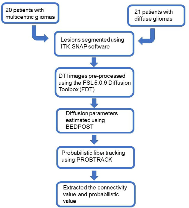
Overview of the image processing pipeline.
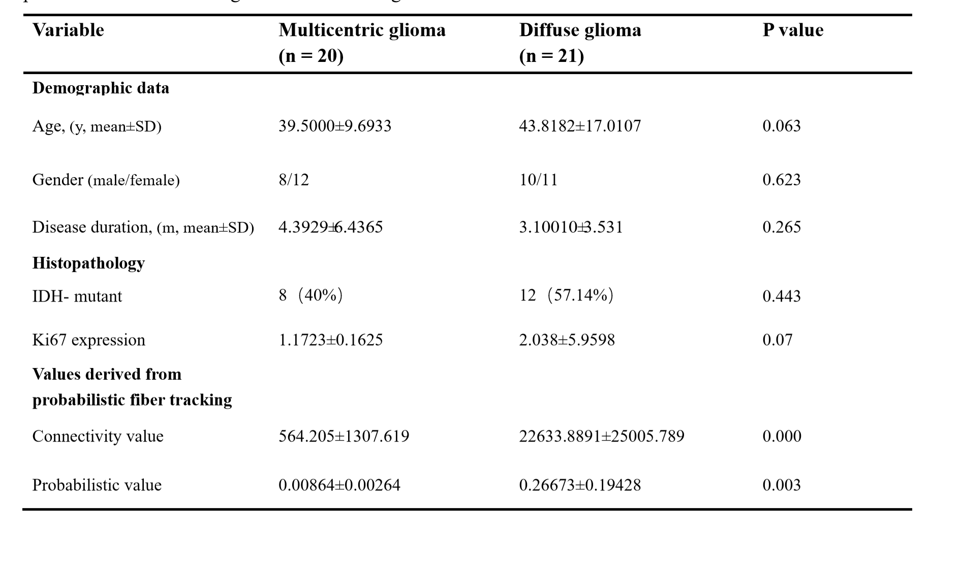
Comparison of demographic, clinical data and values derived from probabilistic fiber tracking by diffusion tensor imaging between patients with multicentric gliomas and diffuse gliomas.
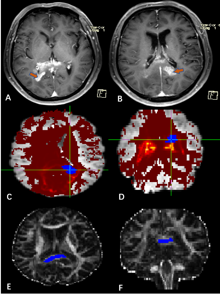
Diffuse glioma. Two lesions (orange arrow) (A,B). The white matter connections between two lesions(red region)(C.D).The likelihood of the probabilistic path (blue region) )(E.F).
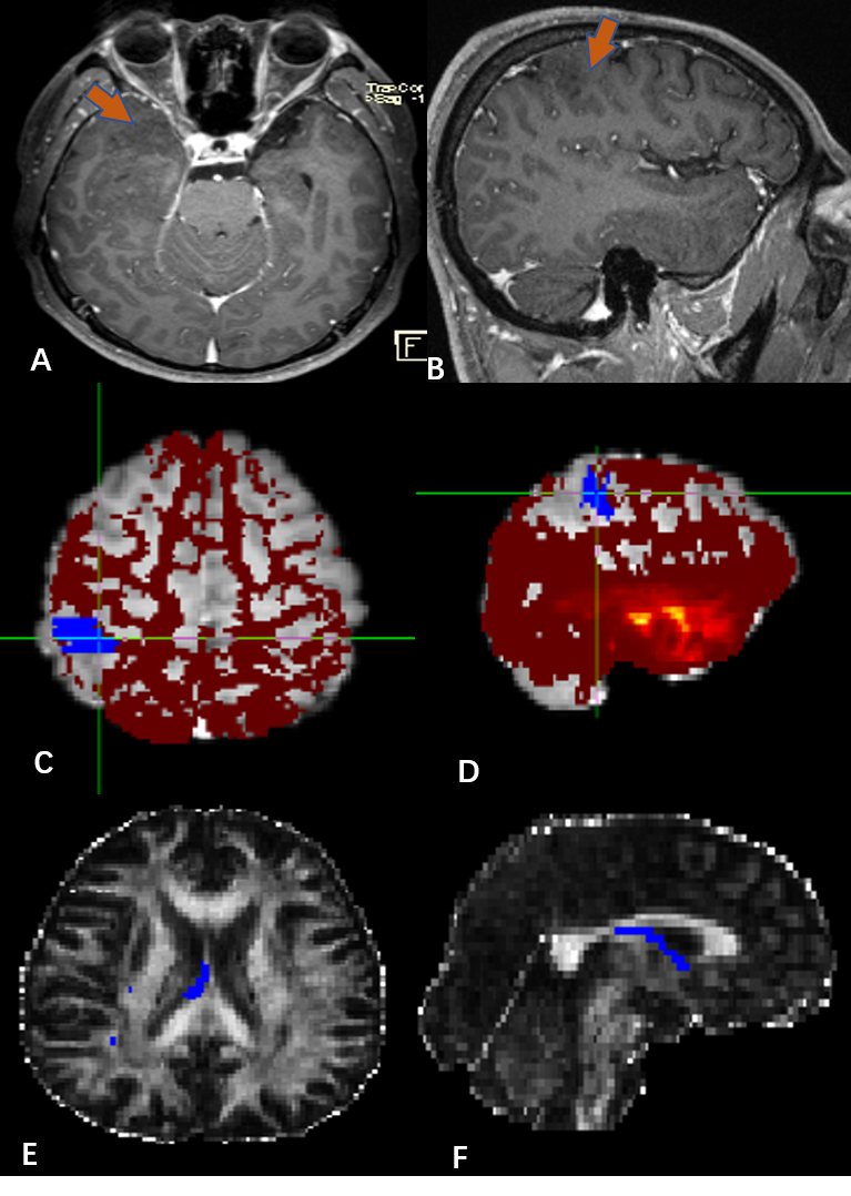
Multicentric glioma. Two lesions (orange arrow) (A,B).The white matter connections between two lesions(red region)(C.D).The likelihood of the probabilistic path (blue region) )(E.F).