1725
A preliminary study on predicting the ACTH&GH hormone immunohistochemistry typing of pituitary macroadenoma based on Radiomics DCE-MRI
Yangyingqiu Liu1, Yanwei Miao1, Ailian Liu1, Qingwei Song1, and Bing Wu2
1Department of Radiology, First Affiliated Hospital of Dalian Medical University, Dalian, China, 2GE healthcare, Beijing, China
1Department of Radiology, First Affiliated Hospital of Dalian Medical University, Dalian, China, 2GE healthcare, Beijing, China
Synopsis
Hormone immunohistochemistry typing of pituitary macroadenomas is assessed by radiomics analysis on Dynamic Contrast Enhanced MRI(DCE-MRI). The results show that DCE-MRI could be used as a new noninvasive method to predictive the hormone immunohistochemistry typingof pituitary macroadenomas.
Purpose
To explore the predictive value of radiomics Dynamic Contrast Enhanced MRI(DCE-MRI) in ACTH&GH hormone immunohistochemistry typing of pituitary macroadenoma.Materials and methods
Fifty patients with histo-pathologically confirmed pituitary macroadenomas were enrolled in this study. Ethical approval and consent forms were obtained. According to the immunostaining of ACTH and GH, they were divided into the ACTH positive (11 patients) and negative (39 patients)group, GH positive (22 patients) and negative (28 patients)group. The preoperative MRI and clinical data were collected. The regions of interest (ROIs) covering the parenchyma of tumor were manually delineated by experienced radiologists, and the radiomics features based on Ktrans, Ve, Kep were automatically generated.Independent t test and Kruskal-Wallis test were used for comparison between groups. ROC curve was constructed to assess the differential ability of independent radiomics features. P<0.05 was considered statistically significant.Results
The placements of ROIs are illustrated in Figure 1.Representative radiomic features of DCE MRI of ACTH and GH groups are shown in Table 1 and Table 2 respectively. In Morphology Metrics features, Compactness(p=0.002) of ACTH positive group was significant decreased compared to negative group, Volume MM(p=0.018)of GH positive group was significant increased compared to negative group. Run Length Nonuniformity(p=0.032) of Ve, Median Intensity(p=0.026), Mean Value(p=0.045), Entropy(p=0.013), MPP(p=0.045), Quantile 25(p=0.021), Quantile 50(p=0.025), Quantile 75(p=0.040), Inertia(p=0.020), Contrast(p=0.019), GLRLM Inverse difference moment(p=0.028) of Kep in ACTH positive group was significant increased, while Energy(p=0.012), GLCM Inverse difference moment(p=0.013), Angular second moment(p=0.007) of Kep in ACTH positive group was significant decreased compared to negative group. Relative deviation(p=0.043) of Ve in GH positive group, Haraentroy(p=0.044), Low grey level run emphasis(p=0.040), Long run low grey level emphasis(p=0.020) of Kep in GH positive group was significant increased compared to negative group. Based on the ROC, the Compactness showed optimal performance for diagnosis(AUC=0.814, sensitivity=89.7% and 72.7%).Discussion and Conclusion
It has been shown by several groups that sparsely granulated somatotroph adenomas and silent corticotroph adenomas are more invasive than other variants [1-2]. In this radiomics DCE MRI based study of pituitary macroadenoma, it was found that the Compactness of ACTH positive group was lower than ACTH negative group,indicating more irregular tumor morphology of the ACTH positive pituitary adenomas; on the other hand, GH positive pituitary adenomas is characterized by proliferation activity, growth rapidly and invasion,which led to larger volumes of as compared to GH negative pituitary adenomas, as supported by the results of this study. Kep in ACTH positive group were generally higher than that in ACTH nagetive group, due to the abundant neovascularization and microvascular permeability in ACTH positive pituitary adenomas. To conclude, Radiomics features of DCE-MRI hold potential value in predicting the hormone immunohistochemistry typing of pituitary macroadenomas.Acknowledgements
No acknowledgement found.References
[1] Kato M, Inoshita N, Sugiyama T, Tani Y, Shichiri M, Sano T et al.(2012) Differential expression of genes related to drug responsiveness between sparsely and densely granulated somatotrophadenomas. Endocr J.2012, 59:221–228.
[2]Jahangiri A, Wagner JR, Pekmezci M, Hiniker A, Chang EF,Kunwar S et al. A comprehensive long-term retrospectiveanalysis of silent corticotrophic adenomas vs hormone-negativeadenomas. Neurosurgery.2013, 73:8–17
Figures
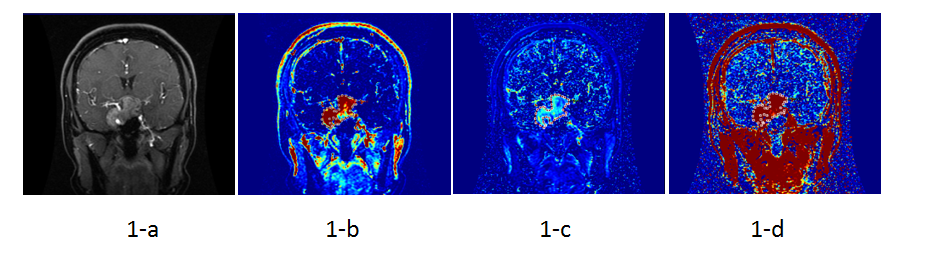
Fig.1(a-d) Images of a 33-year-old
woman with ACTH positive pituitary
adenomas. T1 enhancement image
showing that (Fig.1-a). Fig.1(b-d) showing the ROI were covering the parenchyma
of tumor on Ktrans(Fig.1-b), Kep(Fig.1-c) and Ve(Fig.1-d) maps.
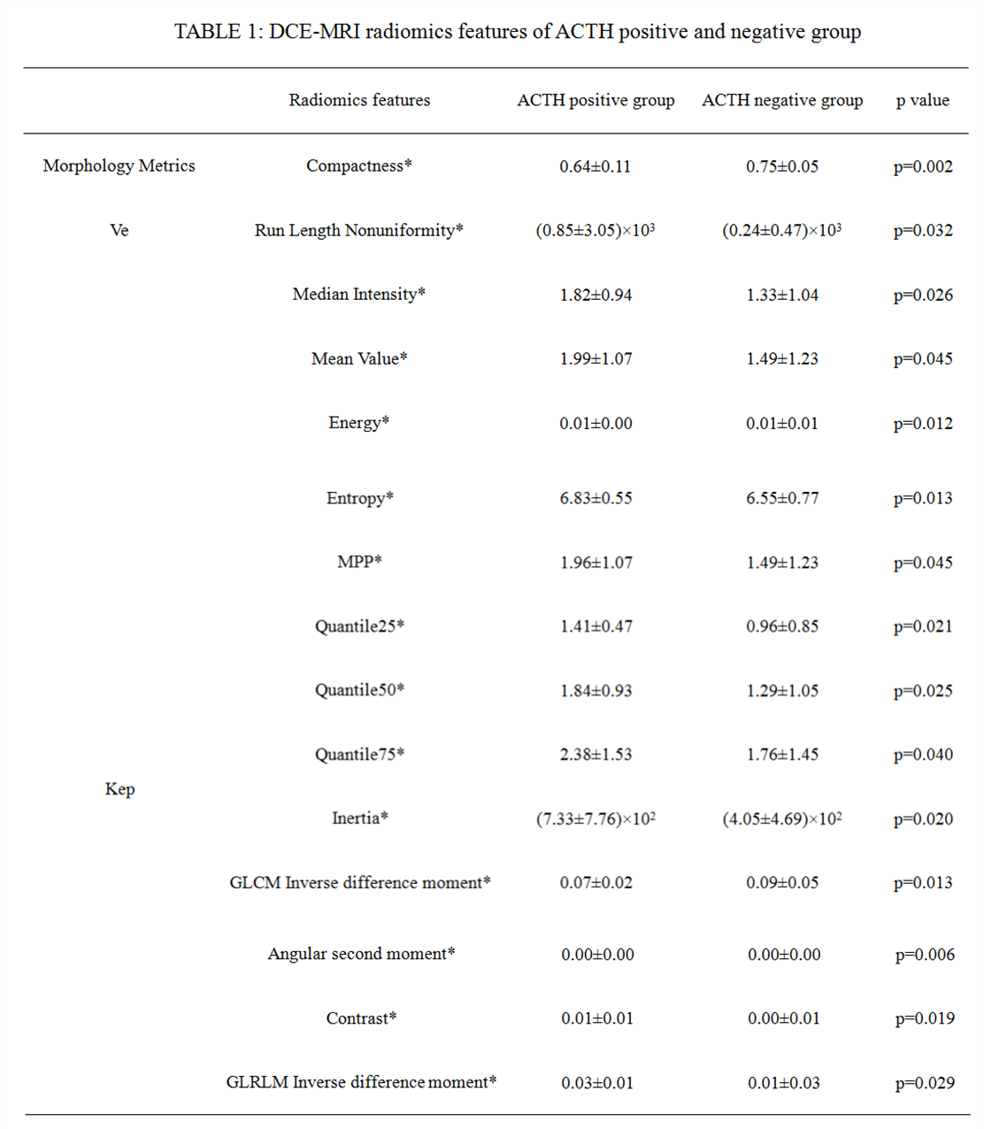
*Data are expressed as mean median±inter-quartile range and was analyzed by Kruskal-Wallis
test
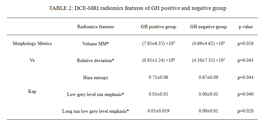
*Data are expressed as mean median±inter-quartile range and was analyzed byKruskal-Wallis
test