1724
Radiomics approach in preoperative differentiation of prolactinoma and non-prolactinoma pituitary adenomas1Henan provincial people's hospital& Zhengzhou University People’s Hospital, Zhengzhou, China, 2Henan provincial people's hospital, Zhengzhou, China, 3Deepwise AI Lab, Beijing, China
Synopsis
The primary treatment for prolactinomas is dopamine agonist, while transsphenoidal surgery is recommended as primary therapy for the non-prolactinomas. This study aimed to evaluate the radiomic model in the differentiation of prolactinoma from non-prolactinomas before surgery. Our results demonstrated that the eXtreme Gradient Boosting model based on 2264 radiomic features extracted from the original and preprocessed sagittal contrast-enhanced T1WI and coronal T2WI yielded the AUC value of 1.000 and 0.828 on the train set and test set, respectively. The radiomic model may have potential for differentiating the prolactinomas from non-prolactinomas, which may be beneficial to guide the treatment plan.
Introduction
Prolactinomas is the most common pituitary adenomas. Dopamine agonist is the primary therapy for prolactinomas, which can rapidly decrease the tumor size in most patients. For non-prolactinoma pituitary adenomas, the primary treatment is generally transsphenoidal surgery. Therefore, the preoperatively identification of prolactinomas is important for implementing effective treatment and improving prognosis. However, conventional magnetic resonance imaging unable to reliably distinguish prolactinomas from non-prolactinoma pituitary adenomas. Radiomics is a promising field that can converts magnetic resonance imaging (MRI) data into a large number of quantitative features. Recently, the radiomics approach has been introduced to extend the study of pituitary adenomas beyond the conventional MRI. The aim of this study was to develop a radiomic model to invasively differentiate prolactinomas from non-prolactinomas before surgery.Methods
This retrospectively study was approved by the local institutional review board. A total of 241 patients with pituitary adenomas who did not receive interventional or medical treatment before the MRI examination and underwent surgical resection after the MRI examination were enrolled from January 2016 to September 2019. The prolactinomas and non-prolactinomas were confirmed by pathological diagnosis. The MRI examination included coronal T2WI and contrast-enhanced sagittal T1WI with a single dose of gadopentetate dimeglumine. The patients were divided into a train cohort (n=192) and a test cohort (n=49). For each MRI series, radiomic features were extracted from the delineated solid component of the tumor of the original images and the preprocessed images, including first-order features, 2D and 3D shape based on features and texture features such as gray level co-occurrence matrix, gray level run length matrix and gray level size zone matrix. The normalized radiomic features were selected by f-test based method to avoid over-fitting. The eXtreme Gradient Boosting model, an ensemble method with decision tree booster was established. Models were built on the coronal T2WI, sagittal contrast-enhanced T1WI and the combination of them. The receiver operating characteristic curve (ROC) was used to represent the performance of the radiomic model in the train set and test set, respectively. The performance was assessed using the area under the ROC curve (AUC), accuracy, sensitivity and specificity.Results
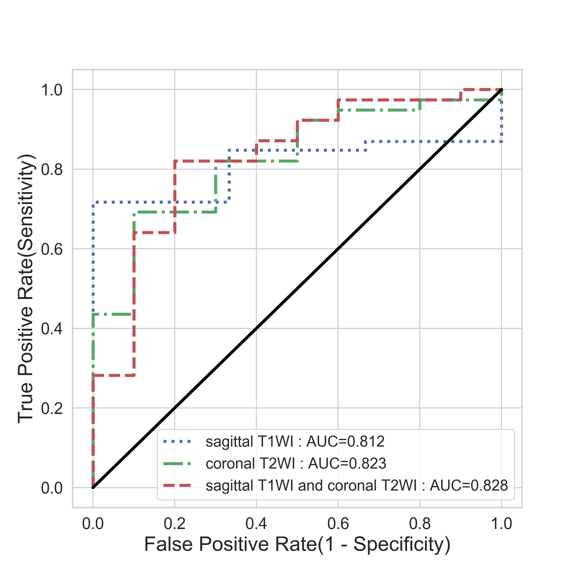
A total of 2264 radiomic features were extracted from the coronal T2WI and sagittal contrast-enhanced T1WI, with 1132 features per series. For the combined mode, 143 significant radiomic features were selected by the p-value threshold of 0.1. The radiomic model based on the selected features yielded the AUC value of 1.000(95% CI 0.981 to 1.000) and 0.828(95% CI 0.693 to 0.921) on the train set and test set, and the accuracy, sensitivity and specificity reached 0.995, 0.994 and 1.000 on the train set, while on the test set, they were 0.796, 0.821and 0.700 respectively. For the models built on the coronal T2WI and sagittal contrast-enhanced T1WI individually, the AUC on the test set reached 0.812(95% CI 0.674 to 0.909) and 0.823(95% CI 0.688 to 0.917) respectively, inferior than the combined model.Discussion
This study established a reliable radiomic model to noninvasively differentiate the prolactinomas from non-prolactinomas. Currently, the serum prolactin level is important for the diagnosis of prolactinomas. However, the pituitary stalk compressed by the tumor or an artifact in the immunoradiometric assay for prolactin can also lead to the prolactin level elevate. Moreover, the conventional MRI has limitations in the differentiation of prolactinomas from non-prolactinomas. The radiomic model with the features extracted from the coronal T2WI and sagittal T1WI may be useful for predicting the prolactinomas and non-prolactinomas before surgery.Conclusion
The radiomic model may have potential for differentiating the prolactinomas from non-prolactinoma pituitary adenomas, which may be beneficial to guide the treatment plan.Acknowledgements
This research was supported by the National Key R&D Program of China (2017YFE0103600), National Natural Science Foundation of China (81720108021, 81601466), and Zhongyuan Thousand Talents Plan Project-- Basic Research Leader Talent (ZYQR201810117).References
1. Molitch ME: Diagnosis and Treatment of Pituitary Adenomas: A Review. JAMA 2017, 317(5):516-524.
2. Klibanski A: Prolactinomas. N Engl J Med 2010, 362:1219-26.
3. Zhang S, Song G, Zang Y, Jia J, Wang C, Li C, Tian J, Dong D, Zhang Y: Non-invasive radiomics approach potentially predicts non-functioning pituitary adenomas subtypes before surgery. Eur Radiol 2018, 28(9):3692-3701.
Figures