1721
Radiomics features of magnetic resonance images as novel predictive factors of bone invasion in meningiomas1Radiology, Lanzhou University Second Hospital, Lanzhou, China, 2Philips Healthcare, Shanghai, Shanghai, China
Synopsis
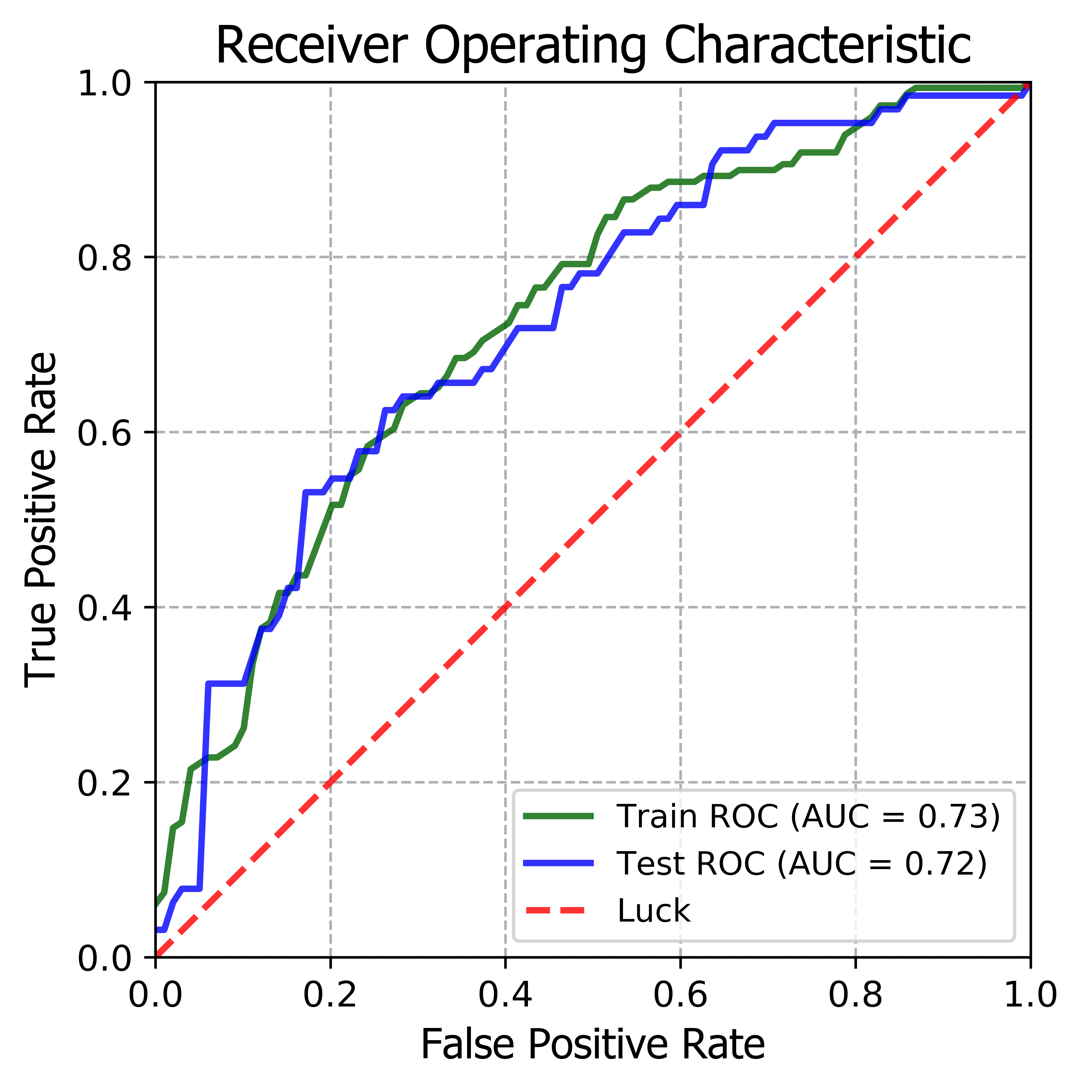
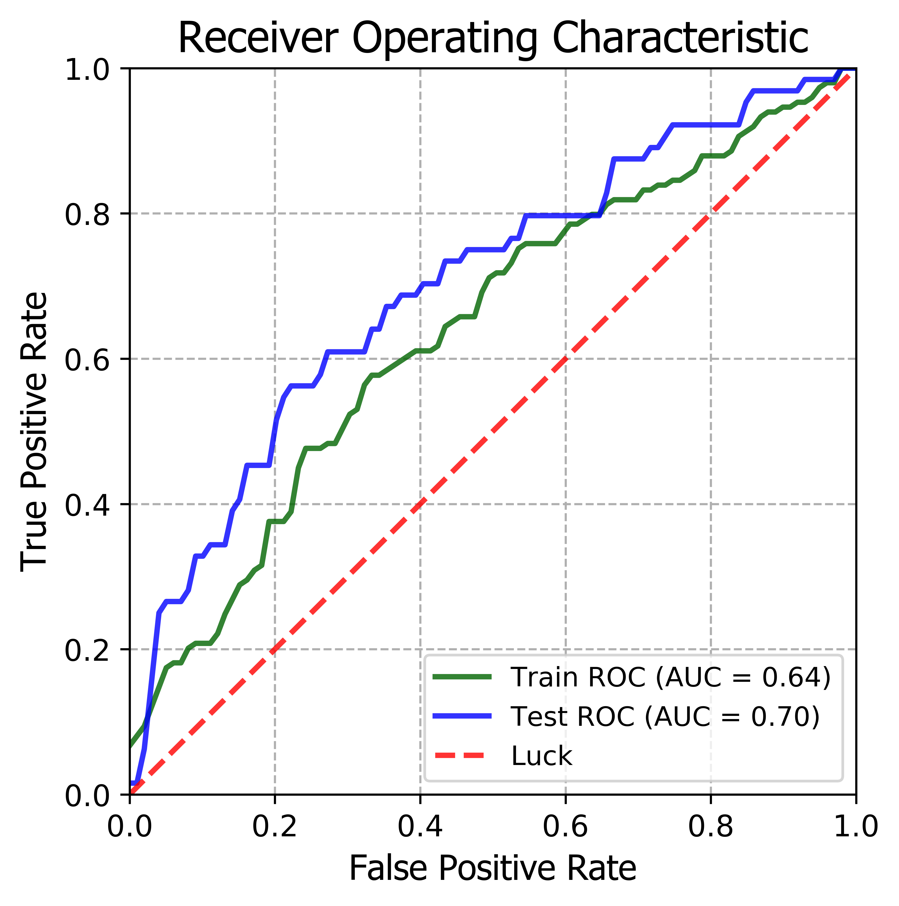
Objectives Radiomics method was used to predict bone invasion. Methods 1227 quantitative imaging features were extracted. Recursive Feature Elimination (RFE) was performed to select the most informative features. Ridge Classifier was chosen to predict model. Results The AUCof the radiomics model derived from CET1WI and T2WI sequence were0.72,0.72and 0.72,0.64 in the training and test datasets , respectively, and combined CET1WI and T2WI sequences were 0.73and 0.72 when predict bone invasion. Conclusions The radiomics model developed in this study may aid neurosurgeons in the pre-operative prediction of bone invasion by meningiomas ,which can contribute to make clinical strategies and predict prognosis.
Introduction
Meningiomas are the most common primary intracranial tumors inadults, accounting for 36.7% of all intracranial tumors1.Bone invasion of meningiomas has been reported in 20%–68% of studies with histopathologically confirmed data2.In such cases, bone hyperplasia or bone infiltration were always caused, even infiltration into adjacent nerves and soft tissuesstructure3,4. Bone invasion is not factored into the WHO criterion for grading of meningiomas, however, the degree of bone invasion and whether it invades bone is a major concern in meningioma surgery, since it can affect directly on the clinical behavior of meningiomas. More importantly, it is predictive of outcome of the patients, including recurrence of cranial involvement, morbidity, and mortality2,4.Bone invasion mainly cause hyperostosis,but bone infiltration has been reported only in10%–40% of cases without hyperostosis, thus making imaging insufficient to predict boneinvasion2,5.Therefore , microscopic quantitative analysis is urgently needed to improve sensitivity of bone invasion. To this aim, radiomics is a novel tool which has emerged in the field of medical imaging analysis in recent years. There have been several applications of radiomics in meningiomas, such as prediction of the grade and differentiation of histological subtypes in meningiomas1,6, prediction recurrence-free survival in meningiomas7. These studies show the value of radiomics in meningiomas, which can also be a potential method for prediction of bone invasion in meningiomas on MR imaging.To the best of our knowledge, until now there is no reported study predicting bone invasion in meningiomas based on the radiomic or texture features analysis. Therefore, in this study, we developed and validated a radiomics model to show the potential association between radiomic signatures and bone invasion in meningiomas.Methods
From January2014 to April 2019,a total of 490 patients diagnosed with pathologically confirmed meningiomas with WHOI grade(448cases),II grade(38cases) and III grade(4cases) were enrolled in this retrospective study(training dataset: n =343; test dataset: n =147). From CE-T1 and T2 MR images, 1227 quantitative imaging features were extracted. Recursive Feature Elimination (RFE) was performed in order to select the most informative features. Subsequently, in training dataset, 5-fold cross-validation was used to compare the different classification algorithms according to the performance of accuracy. Ridge Classifier was chosen to train a predictive model and its performance was further evaluated in the test dataset.Results
Twenty imaging features were selected, which was significantly associated with prediction of bone invasion in both the training and test datasets. The area under the curve (AUC)of the radiomics model derived from CET1WI sequence were0.72and 0.72 in the training and test datasets , respectively. The aucs of the radiomics model had not improved significantly when adding features fromT2WI sequence (two sequences model with AUC of 0.73and 0.72in the training and test datasets, respectively). A radiomics model built from T2WI image features achieved AUCs of 0.72, 0.64 in the training and test datasets, respectively when predict bone invasion.Disscussion
Meningiomas, especially in the convex surface of the brain, are closely related to the skull and easily invade bone .It is reported that the lesions in 25%-50% of cases have an influence on surrounding bone, either infiltrative, osteolytic or hyperostostic changes8-10. In this study, bone involvement accounted for approximately 43.5% of all samples. However, assessability of bone involvement is often limited or varies between histopathologic, operative and imaging reports11.To date, meningioma bone invasion has not been preoperatively investigated. In our study, we investigated radiomic analysis based on preoperative MRI to predict bone invasionin patients with meningioma. The results showed multi-sequence (combiningCET1WI and T2WI )model orCET1WI sequence model was good performance of prediction in both the training set and test dataset .This study revealed that there was a significant association between MR radiomics features and bone invasion in meningiomas. Thus, our analysis provides an alter native non-invasive method to assess bone invasion information for patients and clinicians.Conclusions
The radiomics model developed in this study may aid neurosurgeons in the pre-operative prediction of bone invasion by meningiomas (including WHO I grade, II grade and IIIgrade), which can contribute to make clinical strategies and predict prognosis.Acknowledgements
We greatly thank Dr. Jianqing Sun at Philips Healthcare, Shanghai, for technical assistance of laboratory works.References
1.Yae Won Park,Jongmin Oh,Seng Chan You,et al. Radiomics and machine learning may accurately predictthe grade and histological subtype in meningiomas using conventionaland diffusion tensor imaging. European Radiology.2018;https://doi.org/10.1007/s00330-018-5830-3.
2.Alessandro Della Puppa , Oriela Rustemi, Giorgio Gioffrè,et al. Predictive value of intraoperative 5-aminolevulinicacid–induced fluorescence for detecting bone invasion inmeningioma surgery. J Neurosurg.2014;120:840–845.
3.Bikmaz K, Mrak R, Al-Mefty O. Management of boneinvasive,hyperostotic sphenoid wing meningiomas. J Neurosurg.2007;107:905–912.
4.Fateme Salehi , Shahrzad Jalali , Ryan Alkins ,et al. Proteins involved in regulating bone invasion in skullbase meningiomas.Acta Neurochir.2012;DOI 10.1007/s00701-012-1577-9.
5.Goyal N, Kakkar A, Sarkar C,et al. Does bony hyperostosisin intracranial meningioma signify tumor invasion? Aradio-pathologic study.Neurol India.2012;60:50-54.
6.Niu L, Zhou X, Duan C, et al. Differentiationresearches on the Meningioma Subtypes by Radiomics from Contrast enhanced MRI, A preliminarystudy. World Neurosurgery.2019; doi: https://doi.org/10.1016/j.wneu.2019.02.109.
7 Adriana Olar, Lindsey D. Goodman, Khalida M. Wani,et al. A gene expression signature predicts recurrence-free survival in meningioma.Oncotarget, 2018, Vol.9,(No.22), pp: 16087-169098
8.Talacchi A, Corsini F, Gerosa M. Hyperostosing meningiomas of the cranial vaultwith and without tumor mass. Acta Neurochir (Wien). 2011; 153:53-61.
9.Pieper DR, Al-Mefty O, Hanada Y, et al. Hyperostosis associated withmeningioma of the cranial base: secondary changes or tumor invasion.Neurosurgery.1999;44:742-746.
10.Lau BL, Che Othman MI, Fakhri M, et al. Does putting back hyperostotic bone flap in meningioma surgery causestumor recurrence? An observational prospective study. World Neurosurgery.2019;doi: https://doi.org/10.1016/j.wneu.2019.03.183.
11. Kerstin Zwirner, Frank Paulsen , Jens Schittenhelm, et al. Integrative assessment of brain and boneinvasion in meningioma patients. Radiation Oncology.2019;14:132https://doi.org/10.1186/s13014-019-1341-x.
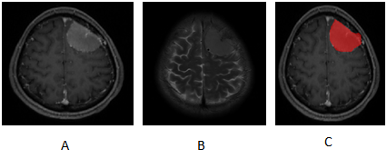
Figures