1709
"ASL-CE mismatch": An Imaging Finding for Diagnosis of Brain Tumor, Multicenter Research1Radiology and Radiation Oncology, Tokushima University Hospital, Tokushima, Japan, 2Tokushima University, Tokushima, Japan, 3Neurosergery, Tokushima University Hospital, Tokushima, Japan
Synopsis
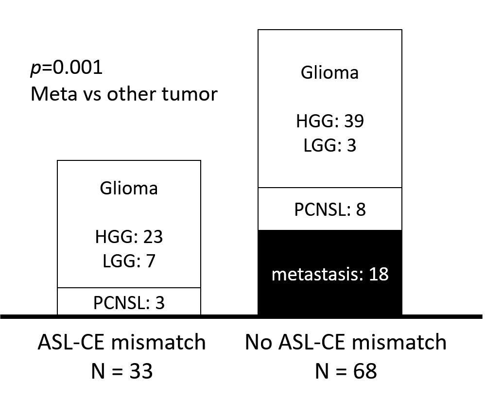
One hundred one cases with brain tumor were included (62: HGG, 10: LGG, 18: metastasis, 11: lymphoma), and divided into two groups by the presence or absence of ASL hyperintensity outside of CE area (ASL-CE mismatch). Only the cases with glioma or lymphoma (23 HGG, 7 LGG, 3 lymphoma) revealed ASL-CE mismatch, and the cases with metastasis didn't (p = 0.001). Similar results were obtained in a sub-analysis where data were divided by MRI vendor. This novel finding may reflect tumor invasion and/or vascular growth in the surrounding brain parenchyma, and is useful for identifying metastasis from the other tumors.
INTRODUCTION
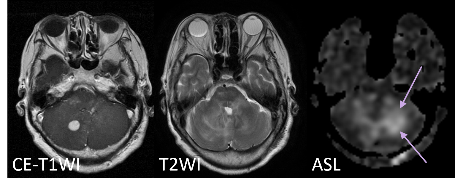
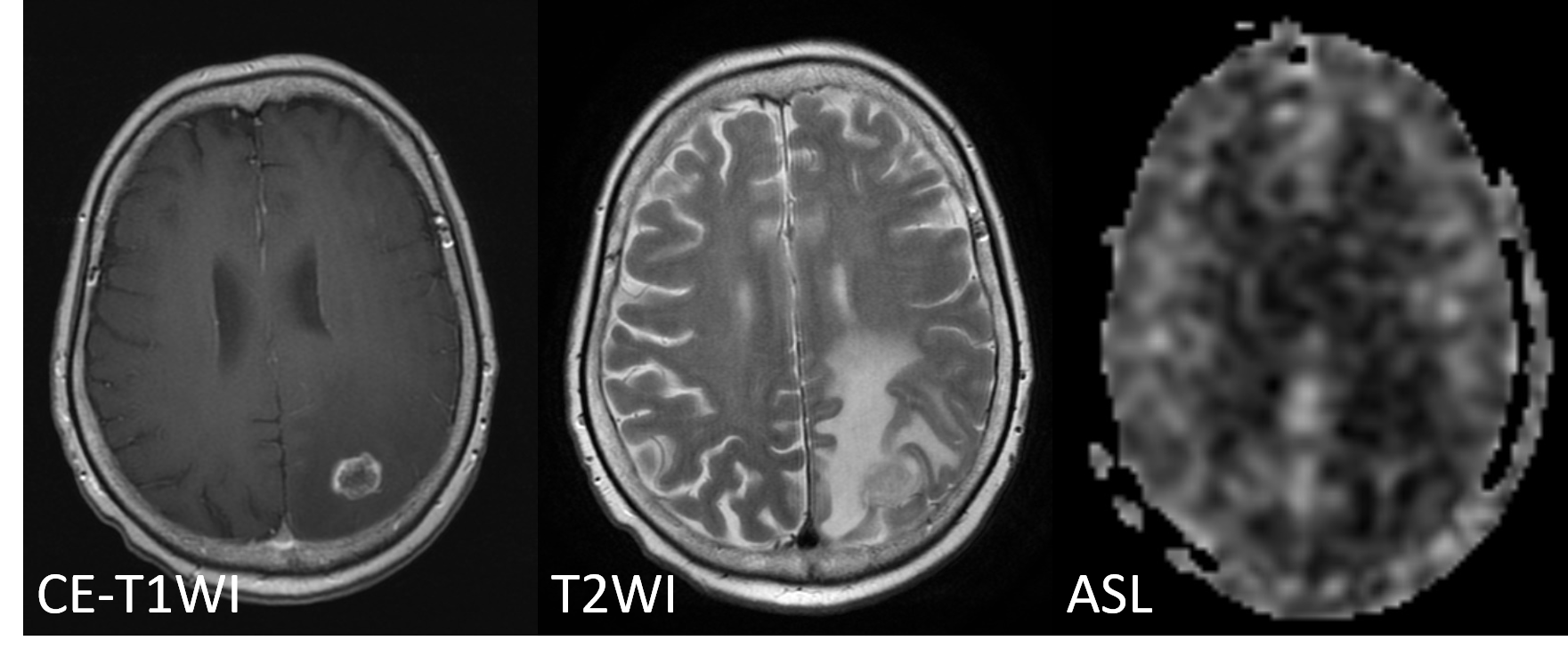
The differential diagnosis of brain tumors is critical in determining optimal therapy and estimating prognosis. High grade glioma (HGG), brain metastasis and lymphoma are common types of brain malignancies in adults and can manifest similar findings on conventional MRI. ASL offers a non-invasive measurement of blood flow image, without any contrast media. We’ve reported the usefulness of ASL hyperintensity outside of CE area (ASL-CE mismatch) for brain tumor diagnosis in single center trial 1 2. At this time, we would validate its usefulness in multi-institutional research.METHODS
One-hundred fifty-four cases who underwent MR examination (T1WI, T2WI, ASL and CE-T1WI) for suspected brain tumor were registered to this institutional review board approved study, and 101 cases were included for the analysis (62: HGG, 10: low grade glioma, LGG, 18: metastasis, 11: lymphoma). The cases were divided into two groups by the presence or absence of ASL hyperintensity outside of contrast enhanced area (ASL-CE mismatch). Tumor histology were compared between the two groups.RESULT
Thirty-three cases showed ASL-CE mismatch (23: HGG, 7: LGG, 3: lymphoma). All cases with metastatic brain tumor didn’t show ASL-CE mismatch, and this finding was useful for differentiate metastasis from the other tumors (p = 0.001, Mann-Whitney U, SPSS v.24). Similar results were obtained in a sub-analysis where data were divided by MRI vendor.DISCUSSION
Metastatic brain tumors tend to be confined to the contrast enhanced area, but gliomas and lymphomas, these are infiltrative tumors that are known to infiltrate outside the contrast enhanced area. The ASL-CE mismatch findings are thought to reflect the tumor invasion outside this contrast area and the accompanying vascular growth. It is also possible that the cytokine produced by the tumor may have induced vascular growth around the contrast zone. Pathological examination is necessary. Pathological examination is needed to confirm what ASL-CE mismatch means.CONCLUSION
ASL-CE mismatch would be potentially useful for diagnosis of brain tumor.Acknowledgements
This work was supported by the research grant from Bayer Yakuhin, Japan.We would like to express our deep appreciation to the participating institutions: Tokushima University, Hokkaido University, Hirosaki University, Tohoku University, Yamagata University, Yamanashi University, Jichi Medical University,Nippon Medical School Chiba Hokusoh Hospital, Kanazawa Medical University, Osaka medical College, Kagoshima University, Japan.
References
1. Abe T, Irahara S, Otomi Y, et al. Discrepancy between arterial spin labeling images and contrast-enhanced images of brain tumors. Proc Intl Soc Mag Reson Med 2015;23:2276 2. Abe T, Mizobuchi Y, Sako W, et al. Clinical Significance of Discrepancy between Arterial Spin Labeling Images and Contrast-enhanced Images in the Diagnosis of Brain Tumors. Magnetic resonance in medical sciences : MRMS : an official journal of Japan Society of Magnetic Resonance in Medicine 2015;14:313-319Figures