1704
Quantitative analysis of MR perfusion imaging in diagnosis and differential diagnosis of alveolar echinococcosis and brain metastases1The First Affiliated Hospital of Xinjiang Medical University, Urumqi, China, Xinjiang, China
Synopsis
Due to similar appearance in conventional MR images, it is often difficult to differentiate cerebral vesicular echinococcosis (CAE) from brain metastases (BM). In this study, user perfusion weighted imaging technique, perfusion characteristics of these 2 diseases were systematically investigated. Our results indicated that significant difference of semi-quantitative parameters relative cerebral blood flow (rCBF) and relative cerebral blood volume (rCBV) from PWI between CAE and BM. Hence PWI can provide valuable additional information in differential disagnosis of alveolar echinococcosis and brain metastases.
Introduction
Cerebral alveolar echinococcosis (CAE) is a zoonotic parasitic disease caused by the larval echinococcosis of echinococcosis granulosus. The magnetic resonance (MR) imaging manifestation of vesicular echinococcosis and brain metastases are quite similar. In practice, it is often difficult to distinguish the two diseases by conventional MR anatomical imaging as well as contrast enhanced imaging. The treatment planning and prognosis evaluation of the two disease are quite different and rely on accurate diagnosis. Hence the differential diagnosis of the two diseases is of high clinical value. Perfusion-weighted imaging (PWI) is a functional imaging technique that has been widely and successfully used in many circumstances. Through the change of PWI metrics, the blood supply and vascular proliferation of the lesion and perilesional tissues can be analyzed to reflect their hemodynamic changes. PWI features many advantages, including easy to perform, nature of semi-quantitative and good contrast between normal tissues and lesions. Therefore, in this study, we systematically investigated the perfusion characteristics derived from PWI of vesicular echinococcosis and brain metastases. Regional perfusion variation of the lesions and differences between the two diseases were also evaluated. By setting up a solid basis, the purpose of our study is to investigate the clinical value of PWI in diagnosis and differential diagnosis of alveolar echinococcosis and brain metastasis.Material and Methods
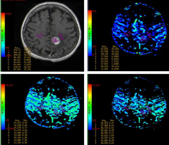
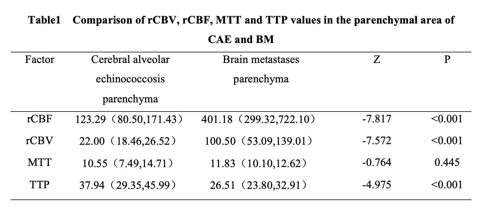
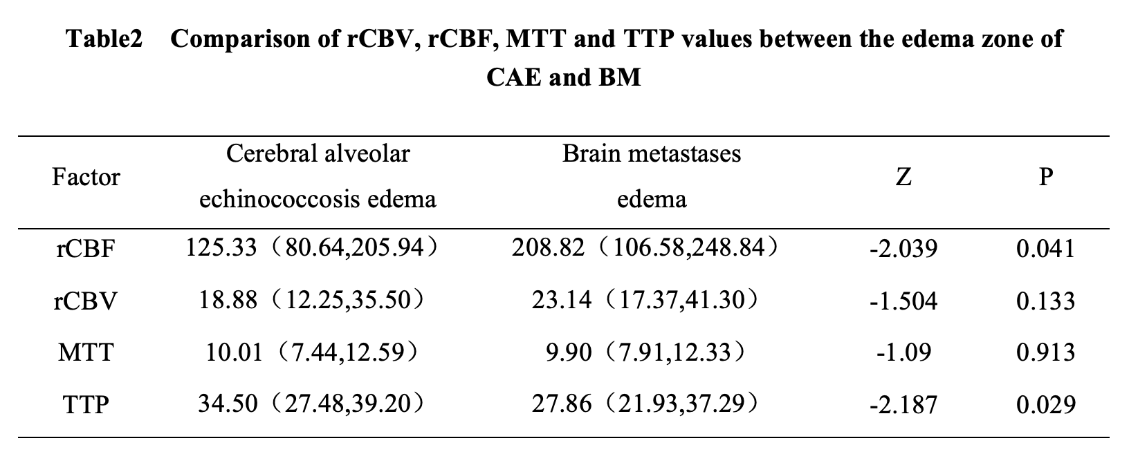
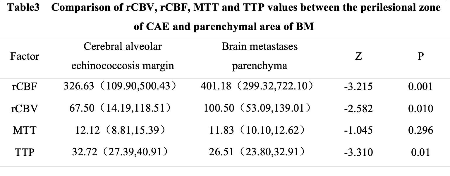
Local ethics committee approved this study and all written informed consents were obtained. Total 10 subjects of alveolar echinococcosis and 10 matched subjects of brain metastasis were enrolled (20 cases, 15 males and 5 females). All subjects underwent MR examination on a 3T scanner (Signa HDx, GE Healthcare, WI, US). Imaging sequences included routine brain anatomical MR scan (axial T1- weighted gradient-echo imaging, axial T2-weighted turbo spin-echo imaging and axial fluid-attenuated inversion recovery imaging), PWI and late contrast enhanced imaging (3 orthogonal plane contrast-enhanced gradient-echo T1-weighted imaging scans). The detailed scanning parameters for PWI are as follows: TR/TE = 1500/14.1ms, flip angle = 90°, Slice thickness = 5 mm, intersection gap = 0 mm, NEX = 1.0, FOV=240×240mm. The semi-quantitative perfusion metrics including relative cerebral blood volume (rCBV), relative cerebral blood flow (rCBF), mean transit time (MTT) and peak time (TTP) were processed on vendor provided advanced workstation (AW4.5, GE Healthcare, WI, US). Measurement was performed with an experienced radiologist with region of interest (ROI) prescribed in the parenchymal area, marginal area and edema area of the lesions for every subjects. The perfusion quantitative parameters of CAE parenchyma, CAE margin, CAE edema, BM parenchyma and BM edema were tested by normality, independence and variance homogeneity test, respectively. The data were inconsistent with normal distribution and uneven variance, and the distribution of data was described by median (25 quartile, 75 quartile), and Kruskal-wallis test was used for statistical analysis. Comparisons of rCBV, rCBF, MTT and TTP values between cerebral vesicular echinococcosis (CAE) and brain metastases (BM) were made with the Mann-Whitney U test. Statistical analysis was calculated in SPSS, Version24.0. P<0.05 was considered significant.Results
For cerebral vesicle lesions echinococcosis and brain metastases, the measured rCBV, rCBF, MTT and TTP in different regions were summarized in Table 1-3.There were significant differences in rCBF, rCBV and TTP between the parenchymal areas of Cerebral alveolar echinococcosis(CAE) lesions and the parenchymal areas of brain metastases(BM). There were significant differences in rCBF and rCBV between the marginal zone of CAE lesions and the parenchymal areas of BE.Discussion and Conclusion
Magnetic resonance perfusion imaging can provide valuable diagnostic basis for the diagnosis and differential diagnosis of Cerebral alveolar echinococcosis (CAE) and brain metastases (BM). According to the results of this study, it was found that the rCBV value and rCBF value of the marginal zone of CAE were lower than those of the parenchyma of BM, and there were statistical differences. The reason was that the growth of BM was accompanied by vascularization, which provided the necessary nutrition and blood for the tumor, resulting in high blood perfusion of the tumor. The marginal band of CAE was slightly hyperper fused due to inflammatory response zone and vascular proliferation, but lower than that of brain metastases. When it is difficult to distinguish BM from CAE disease by conventional nuclear magnetic resonance imaging, Perfusion-weighted imaging (PWI) can be used for differential diagnosis, which can provide reliable basis for clinical practice and make clear the treatment plan.Acknowledgements
No acknowledgement found.References
[1] Yamada K, Wu O, Gonzalez RG, et al. Magnetic resonance perfusion-weighted imaging of acute cerebral infarction: effect of the calculation methods and underlying vasculopathy [J]Stroke,2002,33(1):87-94.
[2] Solomon N, Fields P J, Tamarozzi F, et al. Expert Reliability for the World Health Organization Standardized Ultrasound Classification of Cystic Echinococcosis [J].The American Journal of Tropical Medicine and Hygiene,2017:16-0659.
[3] Stojkovic M, Junghanss T. Cystic and alveolar echinococcosis [J].Handbook of Clinical Neurology,2013,114(114):327-34.
[4] Yang G, Zhang Q, Tang G, et al. Role of Magnetic Resonance Spectroscopy and Susceptibility Weighted Imaging in Cerebral Alveolar Echinococcosis [J].Iranian Journal of Parasitology,2015,10(1):122-127.
[5] Haughton M E, Chan MD, Watabe K, et al. Treatment of brain metastases of lung cancer in the era of precision medicine[J].Frontiers in Bioscience,2016,8(1):219.
Figures