1702
SWI changes in the dentate nucleus in high and low-grade brain stem neoplasms1Radiology, University of Texas Southwestern Medical Center, Dallas, TX, United States, 2Anesthesiology, University of California at Los Angeles, Los Angeles, CA, United States
Synopsis
An investigation that stemmed from observation that there were dentate volume changes on SWI in low grade tumor of the brainstem showed significant decrease in signal intensity in the ipsilateral dentate nucleus in high grade neoplasms and decrease in ipsilateral dentate volumes in low grade neoplasms. We think that dentate iron deposition and its imaging is yet an unexplored field, especially in brain tumor imaging. Further evaluation with QSM and STI with histopathological correlation may also help explore pathways of iron homeostasis.
Introduction
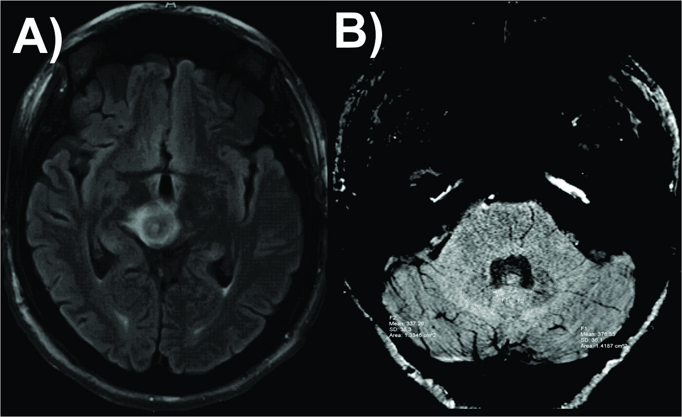
Given their proximity to vital nuclei, brainstem neoplasms present challenges in terms of therapy. Furthermore, management differs between low grade and high-grade neoplasms. While reviewing a case of pontine hemangioblastoma it was observed that the ipsilateral dentate nucleus1-3 was atrophic on SWI images. This led us to question if the differences in SWI signal within the dentate nuclei as a marker of iron content can be used to differentiate high grade and low-grade brain stem tumors.Methods
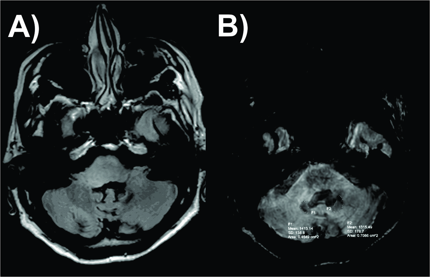
This was an IRB approved, single institution, retrospective study. Primordial was searched for adult patients with pathologic biopsy proven primary or secondary brainstem neoplasms who underwent MR imaging with an SWI sequence from July 2018-June 2019. There were 11 patients (6 females) ranging from 25 to 69-years-old. There were 5 HG (WHO III-IV) and LG tumors (WHO I-II). Age and gender matched controls were selected from a large pool of normal studies done for suspected stroke. The dentate nuclei were identified and manually demarcated on SWI images. Weighted signal intensities were obtained by multiplying signal intensities of dentate nuclei at each slice by the ratio of area of dentate at each slice over total volume of dentate nuclei. Further, average SWI signal intensities were computed by adding weighted signal intensities of each slice (Eqn 1, 2). ROIs were also placed within the normal appearing white matter (NAWM) of the centrum semiovale on both sides for normalization. The average SWI signal intensities for both the dentate nuclei were divided by the NAWM values from respective side. This strategy of weighting and normalization allowed us to compare subjects across scanners/ vendors/ field strengths. Similarly, weighted signal intensity were also obtained for controls.$$ Ave SWI = ∑k Wght SWIk (k= 1 to n).................(Eqn 1)$$
where Ave SWI is average SWI signal intensity, Wght SWIk is weighted SWI signal intensity from the kth slice, n is number of slices where dentate nuclei will appear
$$ Wght SWIk = SIk × (Ark/ Vol)-----------------------(Eqn 2)$$
Where SIk is signal intensity of dentate nuclei from the kth slice, Ark is the area of dentate nuclei at the kth slice, Vol is the volume of dentate nuclei
Results
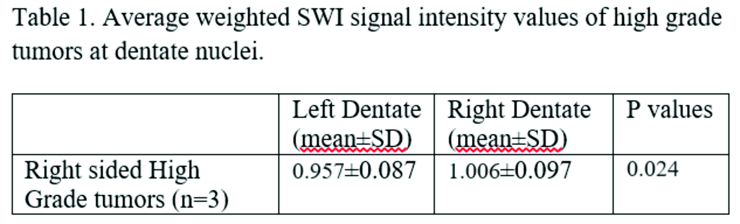
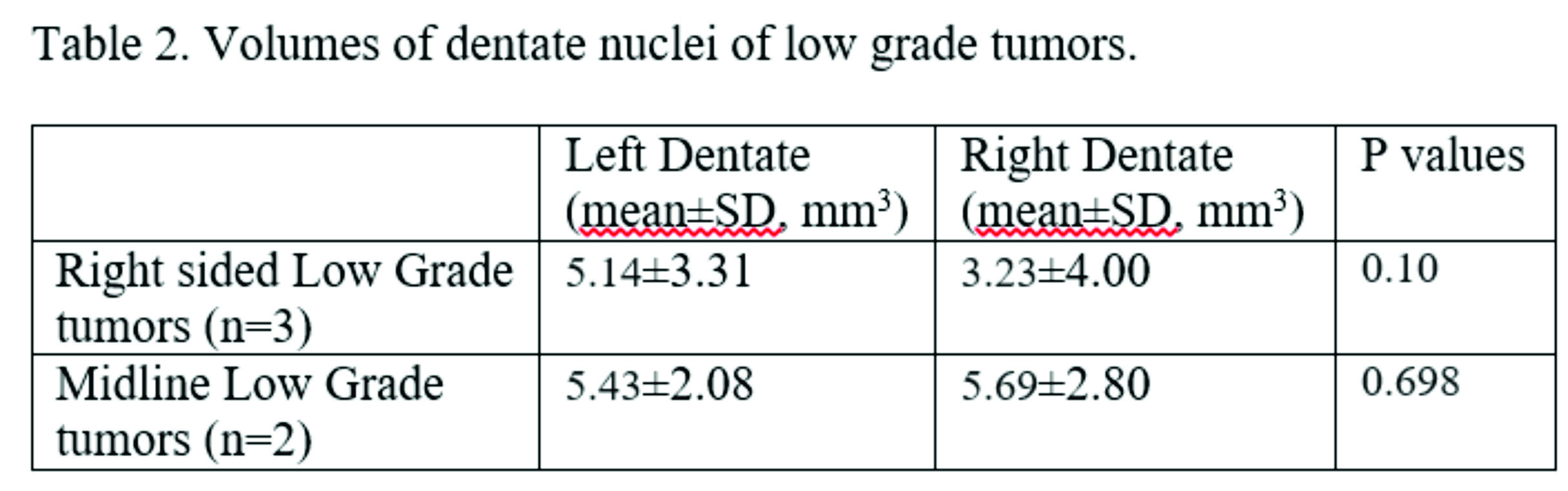
Statistical analysis was performed on SPSS v25. No statistically significant difference was found between the dentate nucleus signal intensity between left and right side for the LG tumor group. For the HG tumor group, a significant difference was found in the dentate nucleus SWI signal (p=0.024), which was increased to the side of the tumor. A similar nonsignificant trend was observed in the LG tumor group (p=0.15). Evaluation of volumes showed no significant difference in the HG group (p=0.451), although there was a trend toward lower volumes of the dentate nucleus ipsilateral to the side of the LG tumor (p=0.10). No significant differences were found in the signal intensities of dentate nuclei of controls.Discussion/ Conclusion
Significant differences in SWI values within the dentate nuclei of high grade tumors, which was increased ipsilateral to the tumor, and may reflect reduced iron content. A trend towards ipsilateral dentate volume reduction as well as increased SWI values was seen in low grade tumors. Although highly speculative it could potentially reflect a yet unexplored pathway for dentate iron homeostasis, possibly intraneuronal. The early destruction of axons caused by high grade tumors vs displacement/ mass effect by low grade tumors may contribute to changes in dentate volume and iron deposition. Future work with the use of advanced susceptibility imaging techniques such as QSM and STI may help to validate these findings.Acknowledgements
References
Bond KM, Brinjikji W, Eckel LJ et al. Dentate Update: Imaging Features of Entities That Affect the Dentate Nucleus AJNR Am J Neuroradiol 2017 38:1467–74
Sang JK, Jae-Hong L, Dae CS Cerebellar .MR Changes in Patients with Olivary Hypertrophic Degeneration AJNR Am J Neuroradiol 1994 15:1715-1719
Phillip GDW, Harding IH, Close TG et al. Longitudinal Evaluation of Iron Concentration and Atrophy in the Dentate Nuclei in Friedreich Ataxia Movement Disorders 2019; 34,3: 335-343
Figures