1690
Dilated PVS affects surrounding white matter non-parenchymal water fraction differently in healthy young and elderly CSVD individuals
Yeerfan Jiaerken1,2 and Zhang Minming1
1Second Affiliated Hospital of Zhejiang University, School Of Medicine, hangzhou, China, 2UNC Chapel Hill, chapel hill, NC, United States
1Second Affiliated Hospital of Zhejiang University, School Of Medicine, hangzhou, China, 2UNC Chapel Hill, chapel hill, NC, United States
Synopsis
We investigated how dilated perivascular space (dPVS) affects water drainage in surrounding tissue and whether there is any distance-related pattern of water distribution around dPVS. We found that The isotropic diffusing water content was higher within dPVS, compared to contralateral normal white matter, and the CSVD subjects had higher within-dPVS water content than healthy subjects, and dPVS showed different distance-related pattern in healthy subjects and subjects with severe CSVD, suggesting dPVS played different roles in healthy subjects and CSVD.
Introductions
To investigate how dilated perivascular space (dPVS) affects water drainage in surrounding tissue and whether there is any distance-related pattern of water distribution around dPVS. Further, we hypothesized that interstitial water clearance processes reflected by dPVS are different in healthy ageing and subjects with severe cerebral small vessel disease (CSVD).Method
28 healthy adult subjects, 20 healthy elderly subjects, and 15 elderly subjects with severe cerebral small vessel disease (lacunar infarction 6 month prior to the scan) were included in our study. DPVS were segmented using a deep-learning based algorithm[1] based on T2 image. Automatically segmented mask were then flipped to the contralateral hemisphere, and overlapping of flipped mask and original dPVS mask were removed from flipped mask, resulting a control mask. Control mask represented normal white matter area where dPVS are highly prevalent. For each voxel, a distance to the nearest dPVS (Dp) and a distance to the nearest control mask (Dc) were calculated. NODDI model was used to obtain the volume fraction of isotropic diffusing water content (Viso, reflecting water with free movement such as water in dilated PVS) and volume fraction of water movement within axon (Vic, reflecting the integrity of myelinated structure) within each voxel. Then we plotted the pattern of NODDI parameter changes with Dp and Dc. [Fig 1.]Results
In all 3 groups, the Vic around dPVS didn’t differ from that of the control points (Pair-wise t-test P values >0.05 at all distance ranges). However, Viso from 0~2mm around dPVS is significantly higher than that of control points in all groups. And Viso from 2~4mm from dPVS is significantly lower than that of control points in healthy adult group and healthy elderly group. In the LA group we didn’t observed such decrease of Viso around dPVS.[Fig 2] ANOVA analysis results suggests that there are significant differences in Viso within 2mm from the dPVS between the 3 groups (P<0.001). Further post-hoc analysis using LSD correction revealed that the differences were significant between each pair of the 3 groups. [Fig 3] The LA group had the highest Viso in within dPVS, while the healthy adult group had the lowest. Pearson correlation test between V in dPVS and the V across the entire normal white matter showed significant positive correlation in the Healthy Elderly group and the Healthy Adult group (P<0.001), but not in the LA group (P=0.888). [Fig 4]Discussion
The discovery of increased Viso at 0~2mm around dPVSs is most likely to be driven by the water signal inside the dPVSs, especially considering our image have a spatial resolution of 2m. Viso in healthy adults and healthy elderly subjects are decreased in areas from 2~4mm around dPVS when compared to normal white matter areas with highly prevalent PVS dilation. This decrease is also visible in subjects with extensive CSVD, but the effect is weaker. One possible explanation is these dPVSs or “MR-visible” PVSs in healthy subjects could accelerate draining of interstitial fluid around it. Though there are currently no direct evidence to prove the dPVSs could accelerate fluid drainage, some studies observed diurnal fluctuation of diffusion signal caused by changes in the perivascular pathway, which suggest the possibility of PVS dilating or closing to adjust fluid drainage. the Viso changes in the dPVS of healthy subjects are highly correlated with the global white matter Viso, which could be interpreted as a mechanism of maintaining fluid balance. While in the CSVD subject the Viso in dPVS and global white matter are no longer correlated in our study, supporting the hypothesis of dysfunctional PVS in CSVD subjects.Conclusion
DPVS showed different distance-related pattern in healthy subjects and subjects with severe CSVD, suggesting dPVS played different roles in healthy and CSVD.Acknowledgements
References
1. Lian C, Zhang J, Liu M et al. Multi-channel multi-scale fully convolutional network for 3D perivascular spaces segmentation in 7T MR images. MED IMAGE ANAL. 2018;46:106-117Figures
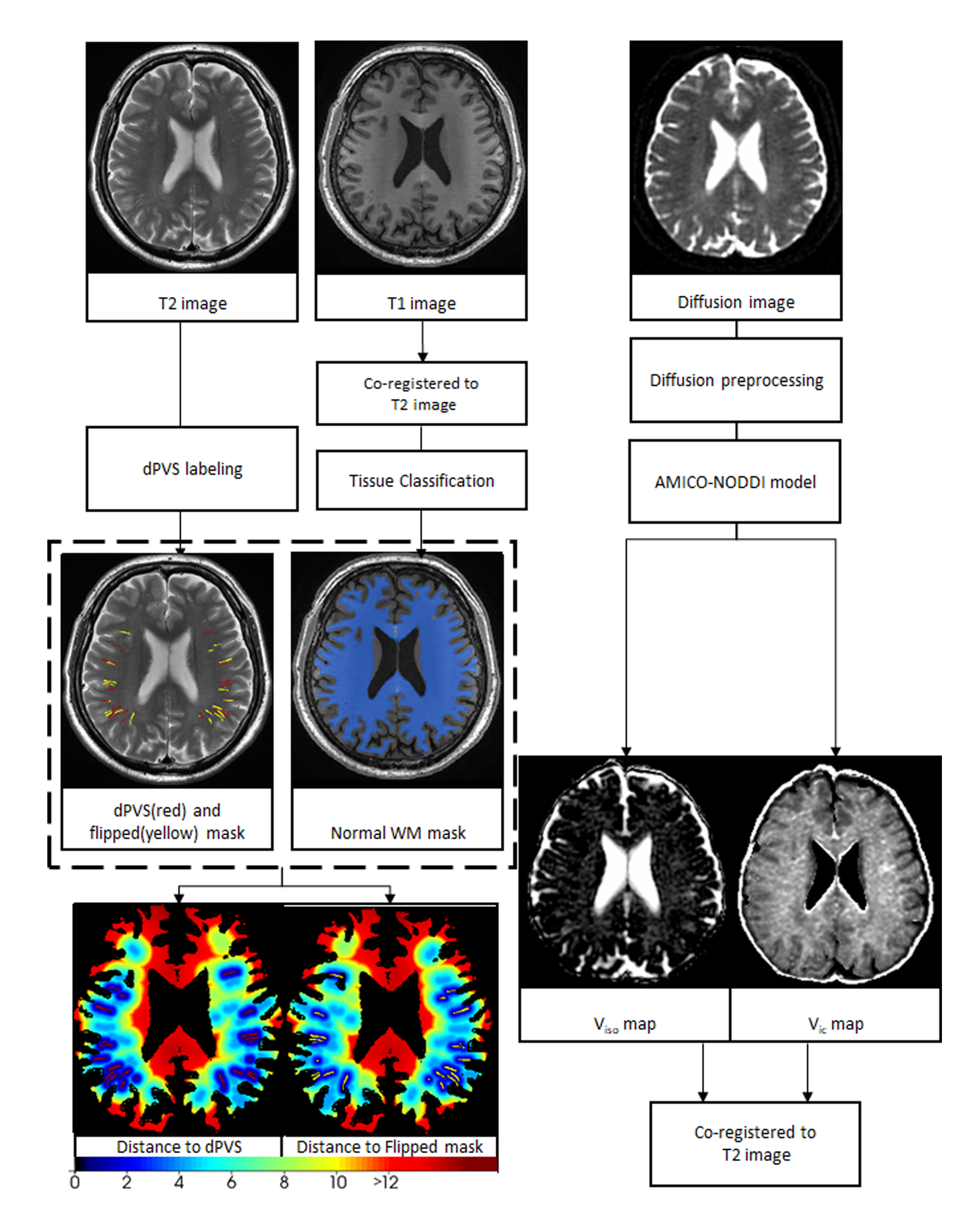
T2 images were used to create the dPVS masks.
Then the dPVS mask were flipped to the contralateral hemisphere, resulting the
flipped mask. The overlapped PVS voxels were removed from the flipped mask.
Then for each voxel on the normal white matter mask, 2 distance values were
calculated: the distance to the nearest dPVS and the distance to the nearest
flipped mask. The respective distance maps were generated by assigning the
distance value to each voxel.
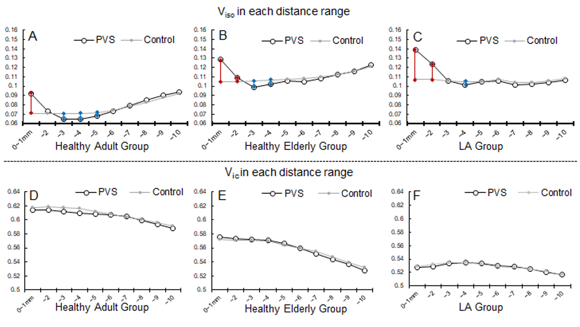
Horizontal axis are the distance to nearest dPVS (or control point). Vertical axis are the mean Viso
(or Vic) in that distance range. E.g. the left most dot
represented the mean Viso (or Vic) value
from all voxels at 0~1mm from a nearest dPVS (or control point), and 2nd
dot represents the range from 1~2mm. Red lines denotes parameters are
significantly higher in the dPVS side than the control side in the connected
pair, and blue line vice versa. P values were adjusted due to multiple
comparisons (10 pairs for each group, 60 pairs in total) based on Bonferroni
method: P<0.05/60≈0.00083.
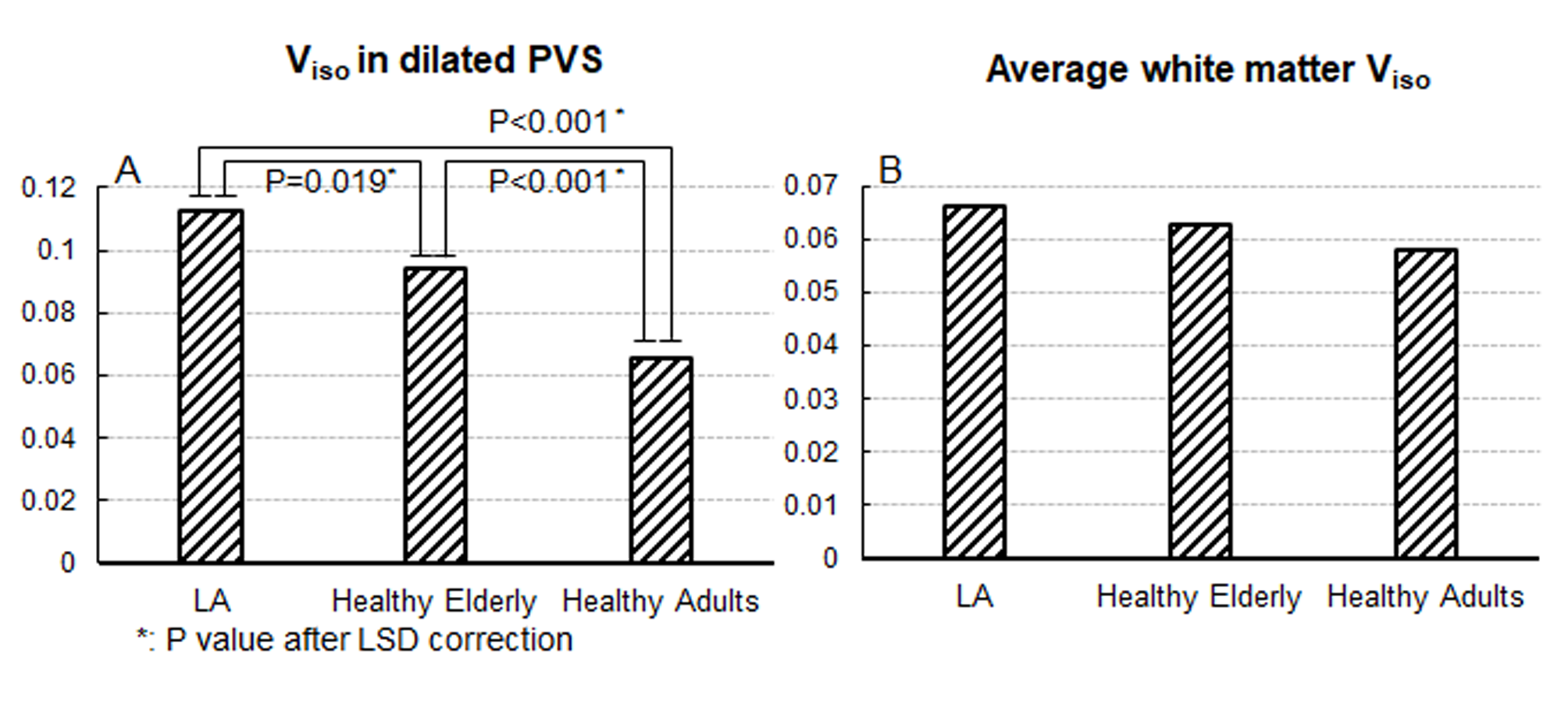
A:
Bar plot showing the differences of Viso in dPVS between
different groups, LA group had the highests had the lowest Viso.
B: the differences of Viso across the entire normal white
matter between the three group. There was a trend of LA group having highest Viso
and healthy adults having the lowest Viso but didn’t reach
statistical significance (ANOVA P=0.241).
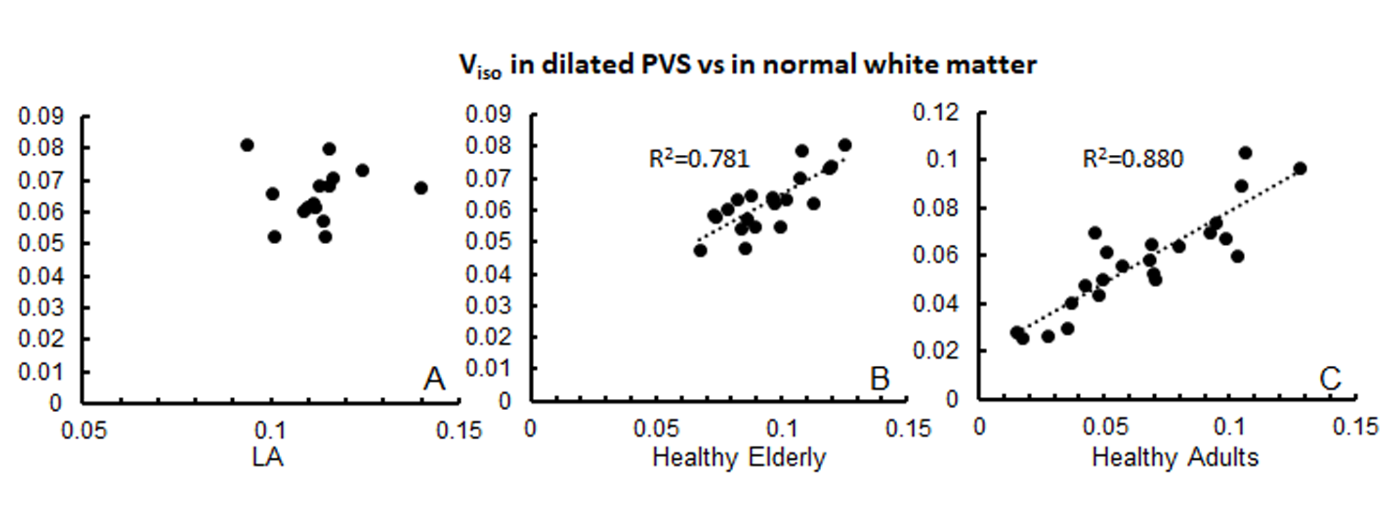
The
Correlation between Viso in dPVS and in normal white matter.
The correlation was not significant in the LA group. Horizontal axes represent Viso
in dPVS and vertical axes represent Viso in normal white
matter.