1673
Abnormality of insular functional connectivity in uremic restless legs:a resting state functional magnetic resonance study1The Second affiliated hospital of Xi'an JiaoTong Uuniversity, Xi'an, China, 2The second affiliated hospital of Xi 'an JiaoTong Uuniversity, Xi'an, China, 3The First affiliated hospital of Neimenggu medical Uuniversity, Hohhot, China, 4The first affiliated hospital of Xi'an JiaoTong Uuniversity, Xi'an, China, 5The second affiliated hospital of Xi'an JiaoTong Uuniversity, Xi'an, China, 6The First affiliated hospital of Xi'an JiaoTong Uuniversity, Xi'an, China
Synopsis
Uremic Restless Leg Syndrome (uRLS) is an uncomfortable complication of inferior limbs and involuntary movements in patients with end-stage renal disease during maintenance of insular, especially during sleep breaks. As an important information processing and processing area, the insular play an important role in sensory, motor metabolism, sensorimotor information processing,and mood regulation. Through this study,We found that the altered morphology of post insular was revealed in uRLS patients, abnormal sensorimotor activities in patients with RLS is closely associated with reduced functional connectivity of insular-S1.Low hemoglobin level was the most important risk factor related to brain functional connectivity feature.
Introduction
Sensorimotor abnormality occurs frequently in patients with end-stage renal disease (ESRD). Approximately 30% of ESRD patients exhibit discomfort sensation such as itch, crawling, aching in the limbs and occur involuntary movement particularly in those who are undergoing hemodialysis [1,2]. This type of sensorimotor abnormality appear or worsen during rest time especially at night, which is the leading cause of poor sleep, decreased quality of life and increased mortality [3]. Several neuroimaging studies suggested that the sensorimotor pathway, subserving sensory modulation and motor activity, is was mediated by the insular [4,5]. However, the pathophysiology of sensorimotor dysfunction in patients with ESRD remains poorly understood.Methods
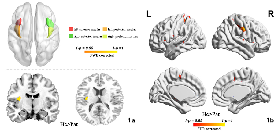
3D high-resolution strcutural imaging was applied in 29 patients with uremic restless legs (uRLS group) and 29 healthy volunteers (control group) with maintenance hemodialysis, The grey matter volume of insular was assessed using a voxel-based morphometry (VBM) analysis.and functional connectivity of insular variation was evaluated by a seed-based correlation analysis, while the seed was choosed as the significant difference of grey matter volume. Spearman correlation analysis was used to explore the significant correlation of functional connectivity and RLS score.The relationship between brain alterations and clinical variables were investigated with Logistic stepwise multiple regression analysis.Results
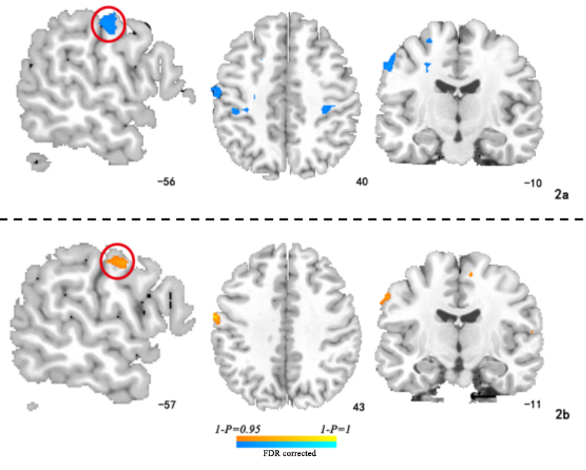
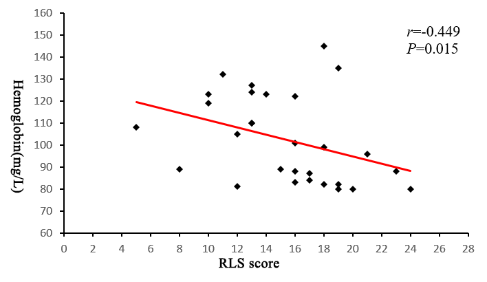
Compared with the control group, the area with reduced gray matter volume in the uRLS group was located on the left posterior insular (P<0.05, FWE correction). Taking the left post insular as a seed region, we further found reduced functional connectivity in patients mainly between the insular and primary sensorimotor cortex,(S1), supplementary motor areas(SMA),and posterior mid-cingulate gyrus(P<0.05,FDR corrected). In uRLS group, the severity score of RLS was negatively correlated with insular-S1 functional connectivity(P<0.05,FDR corrected), while the hemoglobin level was negetively correlated with functional connectivity degree of insular-S1 (r=-0.449 ,P=0.015, FDR corrected).Conclusion
the altered morphology of post insular was revealed in uRLS patients, abnormal sensorimotor activities in patients with RLS is closely associated with reduced functional connectivity of insular-S1.Low hemoglobin level was the most important risk factor related to brain functional connectivity feature.Acknowledgements
Supported
by Major Basic Research Project of Natural Science Foundation of Shaanxi
Province,
China (No.
S2017-ZRJJ-ZDXM-0058) and Clinical Research Award of the First Affiliated
Hospital
of Xi'an Jiaotong
University, China (No. XJTU1AF-CRF-2018-006)
References
[1]Giannaki C D ,Hadjigeorgiou G M , Karatzaferi C , et al. Epidemiology, impact, and treatment options of restless legs syndrome in end-stage renal disease patients: an evidence-based review[J]. Kidney Int, 2014, 85(6):1275-1282.[2] Ding D, Li P, Ma XY, et al. The relationship between putamen-SMA functional connectivity and sensorimotor abnormality in ESRD patients[J]. Brain Imaging Behav,2018, 12(5):1346-1354.[3] Smith SM, Jenkinson M, Woolrich MW, et al. Advances in functional and structural MR image analysis and implementation as FSL[J]. Neuroimage, 2004, 23 Suppl 1: S208-219.[4] Uddin L Q , Nomi J S , Hébert-Seropian, Benjamin, et al. Structure and Function of the Human Insula[J]. Journal of Clinical Neurophysiology, 2017, 34(4):300-306.[5] Craig AD, Chen K, Bandy D, Reiman EM. Thermosensory activation of insular cortex. [J]Nat Neurosci. 2000;3:184–190.[6]Eun Yeon Joo,Jung Sik Kim,Dae Iim Koo,et al. Reduced Cerebral Perfusion of Putamen and Insular Cortex in Patients with Idiopathic Restless Leg Syndrome. J Korean Sleep Res Soc. 2012;9(1):10-14. [7] Berre A P L, Müller-Oehring E M, Schulte T, et al. Deviant Functional Activation and Connectivity of the Right Insula Are Associated With Lack of Awareness of Episodic Memory Impairment in Nonamnesic Alcoholism[J]. Cortex, 2017, 95:15-28.[8] Zhang Y, Yang Y, Bian R, et al. Abnormal Functional Connectivity of Ventral Anterior Insula in Asthmatic Patients with Depression[J].Neural Plasticity, 2017(1):1-11.[9] Lu C , Yang T , Zhao H , et al. Insular Cortex is Critical for the Perception, Modulation, and Chronification of Pain[J]. Neuroscience Bulletin, 2016, 32(2):191-201.[10] Cauda F , D"Agata F , Sacco K , et al. Functional connectivity of the insula in the resting brain[J]. Neuroimage, 2011, 55(1):8-23.[11] An H, Choi B, Son SJ, et al. Renal function affects hippocampal volume and cognition: The role of vascular burden and amyloid deposition[J]. Geriatr Gerontol Int, 2017, 17 (11): 1899-1906.[12] Park HJ, Kim CH, Park ES, et al. Increased GABA-A receptor binding and reduced connectivity at the motor cortex in children with hemiplegic cerebral palsy: a multimodal investigation using 18F-fluoroflumazenil PET, immunohistochemistry, and MR imaging[J]. J Nucl Med, 2013, 54 (8): 1263-1269.[13] Christopher L , Marras C , Duff-Canning S , et al. Combined insular and striatal dopamine dysfunction are associated with executive deficits in Parkinson\"s disease with mild cognitive impairment[J]. Brain, 2014, 137(2):565-575.Figures