1670
White Matter Alterations in Patients with End-Stage Renal Disease: A Diffusion Tensor Imaging Study1Department of Radiology, The First Affiliated Hospital of Dalian Medical University, Dalian, China, 2Philips Healthcare, Beijing, China
Synopsis
End-stage renal disease (ESRD) has become risk factor for vascular disease, stroke and cognitive dysfunction, and the white matter lesions may be associated with these adverse outcomes. Silent white matter alterations may occur in the earlier stages, whereas conventional MR imaging may not be found, This study aim to detect subtle white matter alterations in patients with ESRD through diffusion tensor imaging (DTI). DTI can reflect early occult white matter damage in ESRD patients.
Introduction
End-stage renal disease (ESRD) has become a worldwide public health problem, and it refers to a severe clinical condition with a glomerular filtration rate (GFR) of less than 15 ml/min/1.73 m2 or renal failure and a permanent reduction to 10% of normal status with multiple organ dysfunction1. It is also a risk factor for vascular disease, stroke and cognitive dysfunction, and the white matter lesions may be associated with these adverse outcomes. Therefore, early detection and evaluation of brain structural changes in these patients and intervention therapy have important clinical significance. ESRD results in excessive accumulation of urea and toxic metabolites and hemodialysis is usually performed in patients with ESRD, these all may cause silent white matter alterations in the earlier stages, whereas conventional MR imaging may not be found. Diffusion tensor imaging (DTI) can quantitatively show abnormal pathological changes in white matter microstructure2. Hence, this study aimed to perform diffusion tensor imaging to detect subtle white matter alterations in patients with ESRD.Materials and Methods
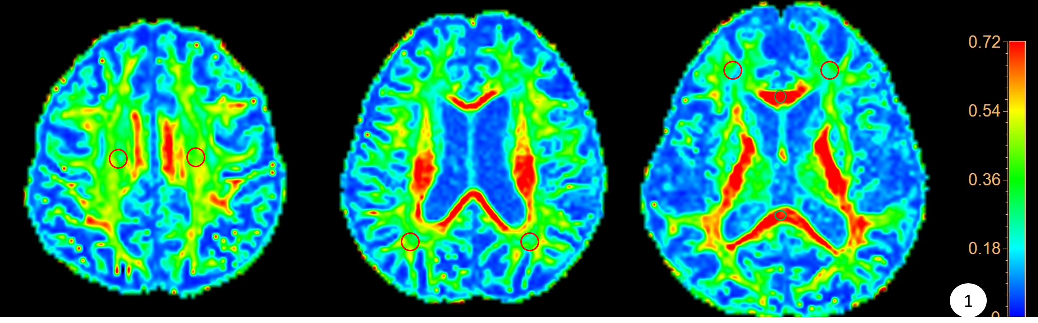
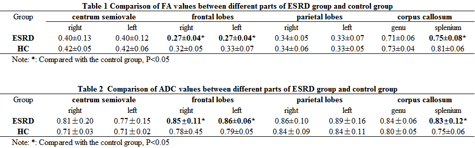
Seventeen maintenance hemodialysis ESRD patients (9 males and 8 females, mean age = 59.1±10.8 years) and 14 age- and sex-matched healthy controls (6 males and 8 females, mean age = 51.9±9.8 years) were participated in this retrospective study. All subjects underwent diffusion tensor imaging (DTI)on a 3.0 T MR scanner (Ingenia CX, Philips Healthcare, Best, the Netherlands) with a 32-channel neck-head array coil. The image is automatically transmitted to the ISP workstation, ROI is placed on the anatomical map to the signal homogenization area, and the size is about 20~30 mm2, Fractional anisotropy(FA), apparent diffusion coefficient(ADC) values were measured symmetrically on bilateral centrum semiovale, frontal and parietal lobes white matter, genu and splenium of corpus callosum. With SPSS 24, T-test was used to analyze significant difference between the two groups. This study has been approved by the local IRB.Results
In patients with ESRD, FA value was significantly decreased in bilateral frontal lobes and splenium of Corpus Callosum WM regions (0.27 ± 0.04, 0.27 ± 0.04, 0.75 ± 0.08) than that in the control group (0.32±0.05, 0.33±0.07, 0.81±0.06), whereas the ADC values were significantly increased (0.85 ± 0.11. 0.86 ± 0.06, 0.83 ± 0.12) vs (0.78 ± 0.45, 0.79 ± 0.05, 0.75 ± 0.06) (P<0.05) (Table 1&2).Discussion
The FA value in the white matter was positively correlated with the integrity of the myelin sheath, the compactness and parallelism of the fibers. The FA value is the most sensitive index reflecting the anisotropic diffusion of water molecules. It is mainly affected by the density, diameter, alignment and the integrity of the myelin sheath. The decrease of FA value indicates the destruction of white matter fiber integrity, density reduction, alignment Consistency is reduced. The ADC value reflects the range and speed of water molecule diffusion. The destruction of cell integrity and the increase of free water content can lead to an increase in ADC value3. The results of this study found that in patients with ESRD, the FA value of the bilateral frontal and corpus callosum decreased and the ADC value increased, suggesting that there are occult lesions in bilateral frontal and corpus callosum in patients with ESRD.Conclusion
DTI can reflect early occult white matter damage in ESRD patients, and the FA value of bilateral frontal lobe and splenium of corpus callosum changes most obviously.Acknowledgements
No acknowledgement found.References
1. Ortiz A, Covic A, Fliser D, et al. Epidemiology, contributors to, and clinical trials of mortality risk in chronic kidney failure. Lancet, 2014, 383(9931): 1831-1843.Chou M C, Ko C H, Hsieh T J, et al.
2. Lee SK, Kim DI, Kim J, et al. Diffusion-tensor MR imaging and fiber tractography: A new method of describing aberrant fiber connections in developmental CNS anomalies. Radiographics, 2005, 25(1): 53-68.
3. A preliminary report of longitudinal white matter alterations in patients with end-stage renal disease: A three-year diffusion tensor imaging study[J]. PloS one, 2019, 14(4): e0215942.
Figures