1668
MRI for studying yoga-induced cortical structural changes in stroke patients1Department of NMR, All India Institute of Medical Sciences, New Delhi, India, 2Department of Neurology, All India Institute of Medical Sciences, New Delhi, India, 3DEPARTMENT OF NMR, ALL INDIA INSTITUTE OF MEDICAL SCIENCES, NEW DELHI, India
Synopsis
This MRI study has evaluated for the first time, the structural changes effected by yoga in post-stroke (ischemic) recovery. Patients (n=6) with deficits practiced Hatha yoga for one hour daily for six months under the supervision of certified yoga trainers. Pre- and 6 months post-yoga intervention assessment involved assessment of NIHSS score and 3D-T1 MRI at 3T MR. Results show individual patients’ positive response to yoga reflected in the significantly reduced NIHSS scores, and in the increased gray matter volume in some areas in brain.
Abstract
IntroductionStroke is not only a leading cause of disability worldwide but also a life-changing event that often leaves the survivors disabled.1 Although physiotherapy is the conventionally used procedure in post-stroke rehabilitation,2 there is an increasing recognition of the positive therapeutic use of meditation and yoga.3,4 The aim of this study was to evaluate using MRI, cortical structural changes in ischemic stroke patients induced by yoga intervention in post-stroke rehabilitation.
Materials and methods
Patient recruitment: The study was carried out with the approval from the Institute Ethical Committee. Six first-ever stroke patients (18-60 years) were enrolled for the study after written informed consent. All the patients had deficits with NIHSS (National Institutes of Health Stroke Scale) score for left hemisphere < 15 and right hemisphere < 10.
Yoga intervention and assessment: The patients were provided Hatha yoga as intervention everyday for one hour for six months under the supervision of certified yoga trainers. NIHSS scores were determined pre- and six months post- yoga intervention.
MR studies: 3D-T1 and 3D-FLAIR acquisition were done pre- and post-yoga intervention at 3T (Ingenia, Philips). Standard FreeSurfer (v6.0) software was used for measuring cortical thickness and volumes using the T1-weighted imaging data. Comparisons between groups (pre- and post-yoga) and changes six months post-yoga intervention with respect to baseline data (pre-intervention) for individual patients were also carried out.
Results
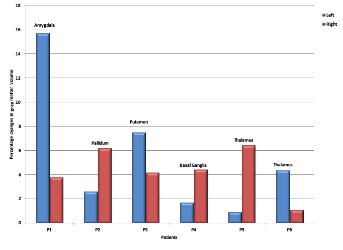
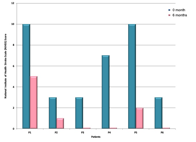
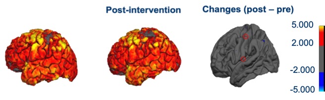
Only data for areas showing significant percentage changes in cortical gray matter volume are shown in Figure 1. It shows the structural changes effected by yoga intervention in cortical gray matter volume for all the patients in both hemispheres. Although the group analysis did not show any significant changes in the gray matter volume, there were interesting structural changes in individual patients. The latter was not reflected in the group analysis probably due to the heterogenous infarct areas. The gray matter volume changes observed are lateralized with significant differences between left and right hemispheres. It is interesting to note that these significant structural changes are observed in structures such as amygdala, thalamus and basal ganglia, which are reported to be affected by meditation and yoga.5 Figure 2 shows the decrease in the NIHSS score for all the patients six months post-yoga intervention reflecting the improvement in the patients. Representative images from a patient showing changes in cortical gray matter between pre- and six months post-yoga are shown in Figure 3. The difference image in the figure has highlighted the areas showing increased gray matter volume.
Conclusion
The societal impact of diseases like stroke is significant since it affects not only the health of individuals but also that of a nation’s economy. There is increasing cognisance of alternative therapies, especially in post-stroke rehabilitation. In this context, the tangible health and therapeutic benefits of yoga alongwith its cost-effectiveness have gained much attention.3,4 With this in focus, the rigors of modern science have been combined with the ancient technique of yoga to understand the changes caused by yoga interventions in ischemic stroke patients. The current MR study has evaluated for the first time, the structural changes in brain due to the effects of yoga intervention in post stroke recovery. The preliminary results have shown the response of individual patients reflected as changes in the cortical gray matter volume. Although the group analysis did not reflect the individual response due to the heterogeneity of the lesions and their locations, the changes observed in individual patients are interesting warranting further in-depth studies and analyses.
Acknowledgements
The work was supported by SATYAM scheme, Department of Science and Technology, Government of India.References
1. World Health Organization, Global Strategy for the Prevention and Control of Noncommunicable Diseases. Report by the Director General. A 53/14 Fift-third World Health Assembly, May 2000.
2.http://www.strokeassociation.org/STROKEORG/LifeAfterStroke/RegainingIndependence/PhysicalChallenges/Post-Stroke-Rehabilitation_UCM_310447_Article.jsp#.W-KLoKeB1Bw, American Stroke Association
3. Jeter PE, Slutsky J, Singh N, Khalsa SBS. Yoga as a therapeutic intervention: a bibiometric analysis of published research studied from 1967 to 2013. J Comp Alt Med, 2015: 1-7, 2015.
4. Sovova E, Cajka V, Pastucha D, et al. Positive effect of yoga on cardiorespiratory fitness: A pilot study. Intl J Yoga. 2015, 8: 134-138.
5. Gotink RA, Vernooij MW, Ikram MA, et al. Meditation and yoga practice are associated with smaller right amygdala volume: the Rotterdam study. Brain Imaging and Behaviour. 2018;12:1631-1639.
Figures