1667
Cerebral Neurovascular Coupling Altered in Patients with Narcolepsy
YuTing Li1
1Department of Radiology, Tangdu Hospital, Fourth Military Medical University, Xian,ShanXi province, China
1Department of Radiology, Tangdu Hospital, Fourth Military Medical University, Xian,ShanXi province, China
Synopsis
Our purpose is to find the cerebral neurovascular coupling altered in patients with narcolepsy.It was reported that Multifunctional MRI can explore the relationship between cerebral vascular perfusion and neural activity. And we found decreased NVC in right and left precentral gyrus, left occipital gyrus, left postcentral gyrus, left fusiform gyrus and right amygdala
Background
Narcolepsy is a chronic neurologic disorder with the abnormal regulation of the sleep-wake cycle, resulting in excessive daytime sleepiness, disturbed nocturnal sleep, sleep paralysis, hypnagogic hallucinations.REM behavior disorder(RBD) is characterized by dream-enacting behavior and impaired motor inhibition during REM sleep. Recent studies have shown that there is a certain relationship between sleep disorders and neurovascular coupling (NVC) dysfunction. However, there is still a lack of clinical evidence for the association between narcolepsy and NVC dysfunction. Multifunctional MRI can explore the relationship between cerebral vascular perfusion and neural activity, thus reflecting NVC condition in people with narcolepsy [1]. Therefore, exploring the relationship between narcolepsy and NVC dysfunction may have important clinical significance for the treatment of narcolepsy.Methods
This study included twenty-seven patients with narcolepsy and twenty-seven healthy controls with no difference in age, gender, BMI and education level. And it was classified as narcolepsy with RBD or without RBD. All participants completed Pittsburgh Sleep Quality Index (PSQI) to assess sleep quality, which shown that narcolepsy patients have lower sleep quality. Magnetic resonance scanner was used to collect the resting Blood oxygen level dependent signal (BOLD), and the resultant Amplitude of low frequency fluctuation (ALFF), fractional Amplitude of low frequency fluctuation (fALFF), dgree centrality (DC) were used to reflect the activity intensity of brain neurons. Cerebral blood flow map (CBF) based on Arterial spin-labeling (ASL) signal was used to reflect cerebral blood perfusion. A two-sample t-test was used for statistical comparison to look for indicators of difference between the sleep disturbance group and the normal group.Results
Through the brain function analysis of the two groups of subjects, we found that the narcolesy patients were abnormal in the PSQI compared with the normal control group. It showed the sleep quality of narcolepsy patients decreased significantly. And we found decreased NVC in right and left precentral gyrus, left occipital gyrus, left postcentral gyrus, left fusiform gyrus and right amygdala . And result of PSQI was matched with MRI.Conclusion
Imaging results showed that there is neurovascular disfunction in narcolepsy patients. Significant correlations were found between the imaging changes of neurovascular uncoupling and the brain function indicators.Acknowledgements
No acknowledgement found.References
[1]. Hu B, Yan LF, Sun Q, Yu Y, Zhang J, Dai YJ, et al. Disturbed neurovascular coupling in type 2 diabetes mellitus patients: Evidence from a comprehensive fMRI analysis. NeuroImage: Clinical, 2019,22: 101802.Figures
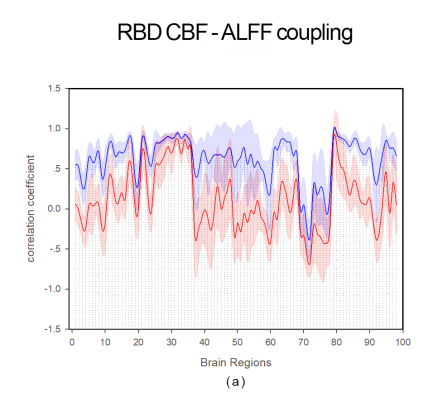
RBD CBF-ALFF Coupling
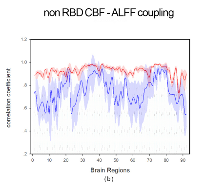
non RBD CBF-ALFF Coupling