1658
Visualizing the Lumen and Wall of Intracranial Artery Stenosis Before and After StentingUsing High Resolution MRI Vessel Wall Imaging
Bing Tian1, Xia Tian2, Zhang Shi2, Qi Liu2, and Jianping Lu2
1Radiology, Changhai hospital, Shanghai, China, 2Changhai hospital of Shanghai, Shanghai, China
1Radiology, Changhai hospital, Shanghai, China, 2Changhai hospital of Shanghai, Shanghai, China
Synopsis
HR-MRI provides important insights of assessing both vessel lumen and wall inintracranial artery stenosis disease.
Purpose:
High resolution MRI vessel wall imaging provides important insights of assessing both vessel lumen and wall in intracranialartery stenosis disease. This study aims to compare the lumen stenosis and wall characteristics change on Vessel wall MRIimaging before and after (different time point) stenting. This would establish the method in evaluating the effect of using thestent for severe symptomatic atherosclerotic intracranial artery stenosis (≥70%).Methods:
This was a retrospective study with patient consent. Thirtytwopatients (20 male, age 55±13) underwent Wingspan stentingtreatment due to intracranial artery stenosis≥70% were included in this study between November 2011 and October 2017.Vessel wall MRI imaging(2D or 3D) was provided before and after stenting (average 354.1 days, range from 1 day to 36months). Patients were divided into two groups(<3 months and ≥ 3 months) according to the time interval of vessel wallimaging after stenting. Pre and poststentingintracranial artery stenosis was measured on postcontrastvessel wall imageaccording to the WASID criterion. Wall characteristics was evaluated including the signal of plaque/vessel wall as well as theplaque/vessel wall enhancement, which were assigned by two neuroradiologists, respectively Plaquetothalamussignalintensity ratio on precontrastT1WI imaging was calculated to represent the signal of plaque/vessel wall. Plaque/vessel wallenhancement was compared using preandpostcontrastT1WI vessel wall images, where the signal intensity of the plaquewas compared with the signal intensity of pituitary. The degree of plaque/wall enhancement was categorized into 3 grades:grade 0 indicates no enhancement, grade 1 represents enhancement less than that of the pituitary infundibulum, and grade 2represents enhancement equal to or greater than that of the pituitary infundibulum. Indeed, degree of stenosis, plaquetothalamussignal intensity ratio, plaque/vessel wall enhancement were compared between preandpoststentingas well as thechange of different groups after stenting.Results:
All patients got preandpoststentingvessel wall MRI imaging. The stent of 26 patients were placed on middle cerebral artery,and 6 were placed on basilar artery. The degree of stenosis was reduced from 86.1% ± 3.2% prestentingto 16.9% ± 6.5%poststenting,which showed good consistency with DSA. The plaquetothalamussignal intensity ratio of poststentingwaslower than that of prestenting.There was no significant difference of stenosis and plaquetothalamussignal intensity ratiobetween different groups. Compared to prestentingimaging, the degree of plaque/wall enhancement showed no significantdifference. But the plaque/wall enhancement score of ≥3 months group was reduced more significant than that of <3 monthsgroup. Agreement between two reviewers were both excellent for the signal of plaque/vessel wall as well as the plaque/vesselwall enhancement.Discussion:
Our studies showed that the degree of stenosis was reduced after stenting, which provide a useful method to demonstrate theeffect of stenting regarding the lumen recanalization in patients with severe symptomatic atherosclerotic intracranial arterystenosis (≥70%). Many studies were performed to analyze the lumen of stenting including CTA and MRA. Previous methodsyielded reliable results of lumen stenosis on poststentingimage due to the stent artifacts. Vessel wall MRI image showed goodconsistency with DSA regarding to stenosis of artery lumen after stenting. Besides the lumen recanalization, vessel wall MRIimaging also can show the wall characteristic changing after stenting. The signal of plaque and enhancement were both theimportant characteristic of atherosclerotic artery disease. Both the plaquetothalamussignal intensity ratio and plaque/vesselwall enhancement were changing significant after 3 months of stent placement, which may provide a direct method to monitorthe plaque dynamic change after stent treatment.Acknowledgements
NoneReferences
[1] Abe A, Sekine T, Sakamoto Y, et al. ContrastEnhancedHighResolutionMRI for Evaluating Time Course Changes in MiddleCerebral Artery Plaques. J Nippon Med Sch. 2018;85(1):2833.
[2] Shi M, Wang S, Zhou H, et al. Wingspan stenting of symptomatic middle cerebral artery stenosis and perioperativeevaluation using highresolution3 Tesla MRI. J Clin Neurosci. 2012 Jun;19(6):9124.
Figures
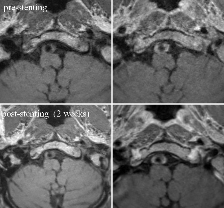
Figure 1. A patient with basilar artery stenosis. Prestentingimage show circle thickness of the vessel and sever stenosis of thelumen. The enhancement score was 3 on prestentingimage. The stenosis, plaquetothalamussignal intensity ratio, andenhancement score didn’t change on poststentingimage after 2 weeks.
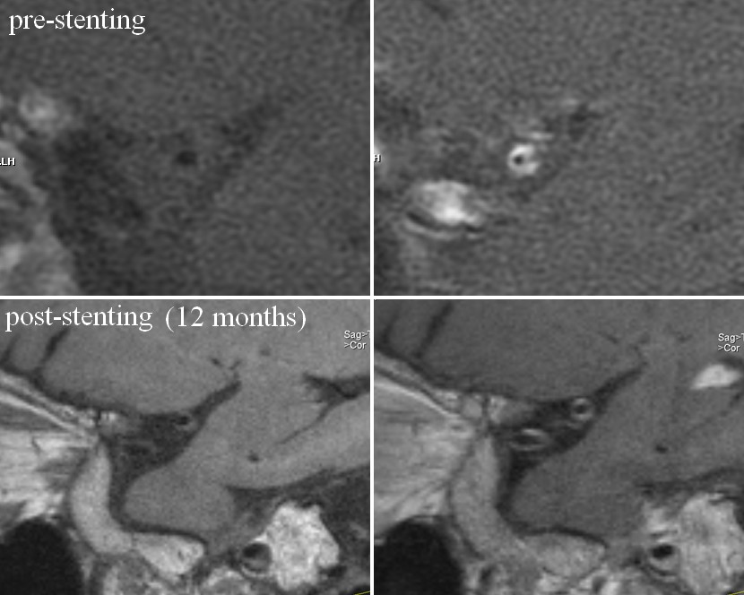
Figure 2. A patient with right middle cerebral artery stenosis. Prestentingimage show eccentricity thickness of the vessel andsever stenosis of the lumen. The enhancement score was 3 on prestentingimage. Vessel wall imaging was performed on 18months after stenting The stenosis degree was lower and the enhancement score was 2 after stenting.