1646
Physiotherapy-induced changes in brain in post-stroke rehabilitation : a longitudinal MRI study1Department of NMR, All India Institute of Medical Sciences, New Delhi, India, 2Department of Neurology, All India Institute of Medical Sciences, New Delhi, India
Synopsis
This MRI based study has evaluated for the first time, the cortical structural changes effected by physiotherapy in post-stroke (ischemic) recovery. Patients (n=7) with deficits did physiotherapy for one hour daily for six months under bi-weekly supervision of certified physiotherapists. Pre- and post-physiotherapy intervention assessment involved NIHSS and mRS scores and MRI at 3T. The latter included 3D-T1, 3D-FLAIR and fMRI (motor task). Preliminary findings showed individual patients’ positive response to physiotherapy reflected in the significantly reduced NIHSS and increased mRS scores, the recovery of fMRI activation and increase in cortical gray matter volume in specific anatomical locations in brain.
Abstract
IntroductionStroke is a leading cause of disability worldwide affecting countries both in terms of economy and work productivity1. Although there are several modalities used in stroke rehabilitation, physiotherapy remains the conventional method2. In this study on ischemic stroke patients, the effects of physiotherapy in post-stroke rehabilitation have been evaluated in brain using Magnetic Resonance imaging (MRI).
Materials and methods
Patient recruitment: The study was carried out with approval from the Institute Ethical Committee. Seven first-ever stroke patients (18-55 years) were enrolled for the study after obtaining written informed consent. All the patients had deficits with NIHSS (National Institutes of Health Stroke Scale) score for left hemisphere < 15 and right hemisphere < 10. The affected areas included parietal and frontal cortex, thalamus, caudate nucleus and putamen.
Physiotherapy intervention and assessment: Standard physiotherapy procedures such as strengthening, balance and coordination exercises, functional and cognitive training, and sensory integration3 were performed by the patient for nearly an hour every day for six consecutive months (twice a week under the supervision of a qualified physiotherapist). NIHSS and mRS (Modified Ranking Scale) scores were determined pre- and six months post-intervention.
MR studies: Longitudinal MRI scans were taken at two different time points, pre- and 6 months post-physiotherapy. Each patient underwent the following studies at 3T (Ingenia, Philips): 3D-T1, 3D-FLAIR and fMRI. The fMRI studies involved both unilateral (motor deficit hand) and bilateral simple motor task of opening and closing of fist. Single-shot EPI sequence was used for the BOLD study with the following parameters: slices - 35; thickness - 4.5 mm; TR/TE - 2000/30 ms; FOV - 230 mm; number of dynamics - 180. The fMRI data was processed with Conn of SPM 12 tool with MATLAB 2018b. T1-weighted anatomical imaging data was used for measuring cortical thickness and volumes using a standard FreeSurfer (v6.0) software. Comparisons between groups (pre- and post-physiotherapy) and longitudinal changes with respect to baseline data (pre-intervention) for individual patients were carried out.
Results
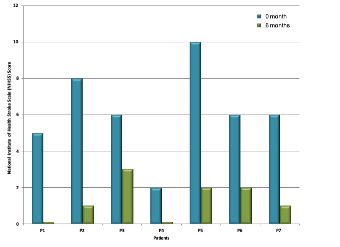
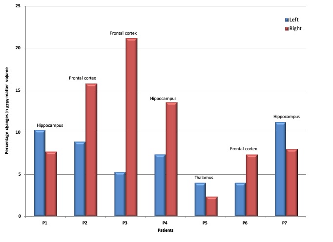
Figure 1 shows the decrease in the NIHSS score for all the patients after six months of physiotherapy. The decrease in the score indicates the degree of recovery. Although all patients’ recovery was reflected in the reduced NIHSS and increased mRS scores, and in the regaining of activation in the motor cortex post-physiotherapy, paired t-test of fMRI data in patients with motor deficit did not reveal any significant changes in activation between pre- and post-intervention for both bilateral and unilateral motor task. Figure 2 shows the physiotherapy induced changes in cortical gray matter volume for all the patients in both hemispheres. The group analysis did not show any significant changes in the gray matter volume. However, interesting structural changes in gray matter volume was observed in individual patients, which was not reflected in the group analysis probably due to variabilities in the region of infarct and other parameters associated with stroke recovery.
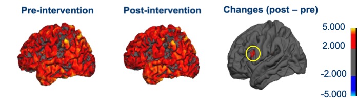
Figure 2 shows the data for areas showing significant percentage changes in cortical gray matter volume. In most of the patients the increase in gray matter volume was observed in the affected area and hemisphere. Figure 3 shows representative images from a patient showing changes in cortical gray matter, pre- and six months post-physiotherapy. The difference image shows the area which had an increase in gray matter volume.
Conclusion
Stroke is a life-changing event that often has a dramatic effect on the survivors. Stroke recovery is influenced by a number of intrinsic and extrinsic factors that also impact the degree of neurological reorganization. Physiotherapy is the conventional method for rehabilitation of stroke patients. Although there are reports on physiotherapy induced changes in physiological functions,4 to the best of our knowledge there are no studies using MRI to assess the structural changes effected by physiotherapy in post-stroke recovery. The current study has evaluated for the first time physiotherapy induced cortical changes in post-stroke recovery using MRI. The preliminary results showed response of individual patients reflected in the outcomes. Though further in-depth studies and analyses are underway, the preliminary results have shown that MRI can play a crucial role in understanding and assessing the effects of physiotherapy in brain in post-stroke recovery.
Acknowledgements
The work was supported by SATYAM scheme, Department of Science and Technology, Government of India.References
1. World Health Organization, Global Strategy for the Prevention and Control of Noncommunicable Diseases. Report by the Director General. A 53/14 Fifty-third World Health Assembly, May 2000.
2.http://www.strokeassociation.org/STROKEORG/LifeAfterStroke/RegainingIndependence/PhysicalChallenges/Post-Stroke-Rehabilitation_UCM_310447_Article.jsp#.W-KLoKeB1Bw, American Stroke Association
3. Donaldson C, Tallis RC, Pomeroy VM. A treatment schedule of conventional physical therapy provided to enhance upper limb sensory motor recovery after stroke: expert criterion validity and inta-rater reliablity. Physiotherapy, 2009; 95: 110-119.
4. Carvalho R, Azevedo E, Marques P, Dias N, Cerqueira JJ. Physiotherapy based on problem-solving in upper limb function and neuroplasticity in chronic stroke patients: A case series. J Eval Clin Pract. 2018; 3: 552-560.
Figures